What do you know about the anatomy of tears formations
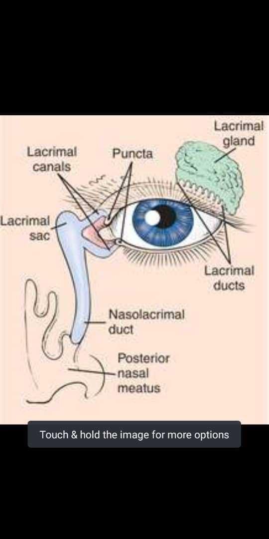
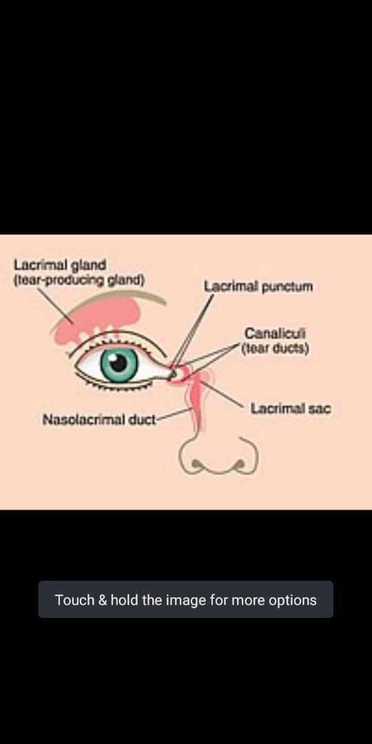
Lacrimal Gland
This gland is about the size of an almond, and sits within the lacrimal fossa, located in the superior and outer edge of the orbital roof. The gland is divided into two sections anatomically. These are the small palpebral portion that lies closer to the eye, and the orbital portion that forms around four ducts. These ducts then combine with the 6 ducts of the palpebral portion, and are secreted onto the surface of the eye. The lacrimal gland is composed of cells that produce proteins and electrolytes, and cause water to follow by osmosis.
Lacrimal gland - ventral view
Lacrimal gland - ventral view
The blood supply of the gland is from the lacrimal artery (a branch of the ophthalmic artery), with the venous drainage through the superior ophthalmic vein. The gland is innervated by the parasympathetic lacrimatory nucleus of the facial nerve (which originates in the pons).
Lacrimal artery - lateral-left view
Lacrimal artery - lateral-left view
The facial nerve leaves the skull via the foramen lacerum by hitching a ride with the greater petrosal nerve, eventually reaching the eye by merging with the lacrimal and zygomatic divisions of the ophthalmic division of the trigeminal nerve. The nerve also synapses in the pterygopalatine ganglion. The sympathetic innervation arises from the superior cervical ganglion, before merging to form the deep petrosal nerve, which merges with the greater petrosal nerve.
Lacrimal Canaliculi/Canals
These are small channels that lie in each eyelid, and commence at the puncta lacrimalia; small openings where the tears are drained from the surface of the eye. These canaliculi are divided into the superior duct and the inferior duct that drain into the lacrimal sac. They are lined with stratified squamous epithelium.
Lacrimal canaliculus - ventral view
Lacrimal canaliculus - ventral view
Lacrimal Sac
This is the upper dilated end of the nasolacrimal duct. It connects to the lacrimal canaliculi, which function to drain the tears from the eyes surface to the nasal cavity via the nasolacrimal duct. The cells that line these canaliculi are stratified columnar epithelium, with goblet cells.
Lacrimal sac - ventral view
Lacrimal sac - ventral view
Nasolacrimal Duct
These drains the tears into the nose, and are drained away just anteroinferiorly to the inferior nasal concha. The cells that line this duct are stratified columnar epithelium. The membrane at the end of the tear duct (the valve of Hasner) may fail to open at birth, resulting in obstruction of the duct.
Nasolacrimal duct - ventral view
Nasolacrimal duct - ventral view
Meibomian Glands
Lipids (meibum) are secreted by these specialized sebaceous glands and they form a part of the tear film. There are approximately 50 glands on the upper eyelid, and 25 on the lower lid. These are squeezed from the glands upon blinking, and they have numerous functions including closing the eyelid airtight and also preventing tear spillage onto the cheek, by maintaining the tears between the oiled edge of the eyelid and the eyeball.
Tear Film
The tear film consists of lipids, water and mucins. Mucins are both lipids and long hydrophilic molecules, and are made by goblet cells that are scattered over the surface of the conjunctiva. The tear film is able to maintain a wet layer as it binds to the corneal and conjunctival epithelium, which is coated with a layer of mucins known as the glycocalyx.
Conjunctiva (Superior conjunctival fornix)
Conjunctiva (Superior conjunctival fornix)
Tears wet both the front of the eye, and the inner surface of the upper eyelid. The relatively thin tear layer on the surface of the eye is covered by an oily layer that prevents tears from evaporating. This oily layer of lipids is largely composed of nonpolar lipids secreted by tarsal glands. The function of the lipid layer is dependent on the inner layer of lipids that are composed of both hydrophobic and hydrophilic lipids. As a result, the layer can form an interface with the underlying aqueous fluid. The aqueous layer contains igA antibodies, electrolytes and antibacterial enzymes that protect the eye from infection.
The tear film functions to protect the eye from shear forces during eye movements and blinking, maintains a smooth transparent optical layer and also shield the eye surface from environmental insults. The negative charge of the tear film also functions to repel negatively charged bacteria. When we feel sad, there is a large increase in lacrimal output, which dilutes the tear film. This then results in more watery tears that therefore flow more readily.
Congratulations @tochprince! You have completed the following achievement on the Steem blockchain and have been rewarded with new badge(s) :
You can view your badges on your Steem Board and compare to others on the Steem Ranking
If you no longer want to receive notifications, reply to this comment with the word
STOPVote for @Steemitboard as a witness to get one more award and increased upvotes!