Cardiac tumors: Auricular Myxoma
During the last decade, researchers in the medical field have been very interested in the study of cardiac tumors, as a result of their increased clinical presentations and their lethal complications. His diagnosis was mostly Post Mortem, during the autopsy, but nevertheless thanks to scientific and medical advances have been detected after the same complex studies.
Tumors at the level of the heart are usually infrequent. From the histopathological point of view, approximately 75% are benign and the rest malignant, in the majority of cases they are sarcomas. However, their diagnosis is essential since they generate great fatal complications, such as Superior vena cava syndrome and that most of the treatment is surgical reception.
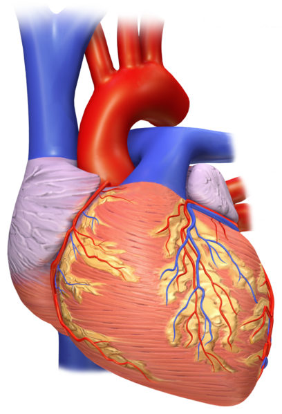
The human heart viewed from the front. Image with free rights of use. CC BY-SA 3.0.

A little history
In the past, the diagnosis of cardiac tumors is made under the study of corpses without presenting a certain cause. It was only until the middle of the sixteenth century that Boneti described the first finding of a cardiac tumor after autopsy. Later in 1934, it was possible to publish a study based on 226 necropsies under Dr. Lymburner, which reports the presence of intracardiac tumors.
Finally only until the mid-twentieth century, which succeeded in executing the successful resection of a cardiac tumor, using the CCE extracorporeal circulation technique developed by Crafoord, for now the pre-Mortem diagnosis has increased thanks to the presence of more advanced medical equipment without However, it continues to be a medical challenge since, due to its rare and scarce presentation, its diagnostic suspicion is poor and often go unnoticed.
Cardiac Anatomy
The heart, noble organ of our organism with an approximate size to a closed fist, with an estimated weight between 250 to 300gr. It is located in the mediastinum, between both lungs, above the diaphragm, behind the sternum and in front of the thoracic spine.
This vital organ fulfills the important function of receiving, pumping and distributing blood to the different tissues of the organism, that is why several literatures describe it as a cardiac pump. Every day our heart generates an average of 100,000 beats, with an approximate pumping of 7,570 liters of blood.
Layers of the heart
Fine pericardium and double membrane that surrounds the heart, whose function is to protect and adhere it to the chest wall. It consists of two named portions; Fibrous pericardium and Serous Pericardium and in the middle of both membranes a virtual space through which a serous fluid called pericardial fluid flows, which reduces friction during cardiac movements.
The wall of this essence organ, is composed by; Epicardium, Myocardium and Endocardium.
It consists of 4 cavities, of which two are superior and are called auricles are wrinkled structures in the form of bags, also known as Orejuelas. While the larger structures are located internally and known as ventricles, the left ventricle is the largest and thickest that this is responsible for pumping blood through the aortic valve and from there to the rest of the human body.
There is a small muscle structure that interposes between the atria and the ventricles and separates them into right and left and this is called Septum or septum.
Heart valves
Essential and important for the good cardiac functioning, since it allows the passage and exit in turn regulate the blood flow in the heart, 4 valves are described and these are:
Tricuspid valve, between the right atrium and right ventricle.
Valve valve, allows the passage of blood flow between the left atrium and left ventricle.
Aortic valve, the largest, and it is the blood passage from the left ventricle to the aorta.
Lung valve, from the right ventricle to the pulmonary arteries. It is responsible for the oxygenation of the blood.
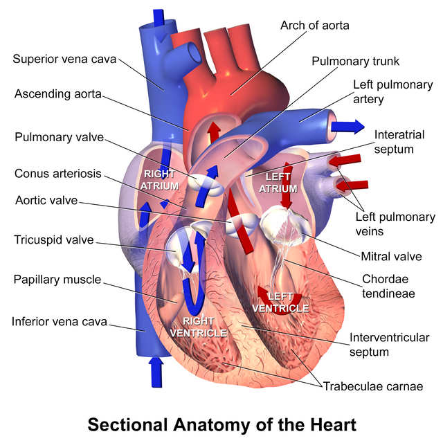
Sectional Anatomy of the Heart with Path of Blood Flow. Image with free rights of use. Author: Blausen Medical Communications, Inc. CC BY 3.0.
Cardiac Tumors
Once having a clear review of the anatomy of the heart, we can enter into matter regarding tumors in the heart, being an infrequent pathology as already established from the beginning.
In general, the tumors are divided into benign and malignant.
Benign primary cardiac tumors
Myxoma.
Rhabdomyoma.
Fibroma.
Hemangioma.
Atrioventricular node.
Granular cells.
Lipoma.
Mesothelioma of node A-V.
Teratoma.
Malignant primary cardiac tumors
Sarcoma.
Lymphoma.
Presentation of the real clinical case
We must note that cardiac tumors can present with multiple cardiac and noncardiac manifestations.
The location and size of the tumor are the factors that will determine the signs and symptoms (some of these symptoms are usually similar to other presentations of heart disease) such as:
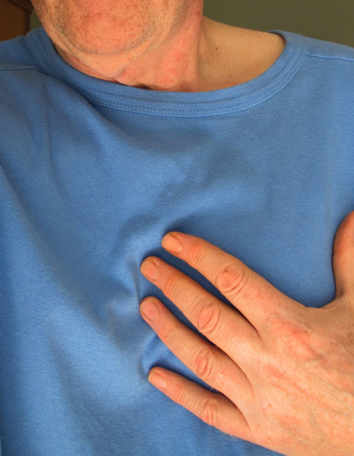
Chest pain. Public domain image. Source Pxhere
Benign tumor: Myxoma
Myxomas are the most frequent primary cardiac tumors in all age groups, representing 75% of the cases found in necropsies, and in almost three quarters of the tumors treated surgically.
They appear at all ages, with a peak incidence between 40 to 60 years, affecting more often the female sex.
The vast majority of cases of this type of pathology occur when lifting, ie a genetic predisposition has not been established, however cases have been reported that have a family character and are transmitted as an autosomal recessive or are part of a syndrome consisting of a complex of anomalies, such as lentigines or pigmented nevi, primary adrenal nodular disease, myxomatous breast fibroadenomas; Testicular tumors, among others.
One of the characteristics of these tumors is that after your resection has the ability to recur and reappear after surgery.
Anatomically this type of lesion is located 75% in the left atrium, 20% in the right atrium and the rest 5% in the ventricles.
Histological characteristics
Myxomas are gelatinous structures formed by myxomatous cells embedded in a stroma with abundant glycosaminoglycans.
All are pedunculated, have a fibrovascular stem and measure 4 to 8 cm in diameter. Most sporadic myxomas are unique and are located in the atria, most often in the left.
Unlike familial myxomas or myxomatous syndrome, they affect younger individuals, are multiple and have a higher recurrence rate after surgical reception.
Clinical Manifestations of the Myxoma
One of the most common that can be seen in this type of cardiac tumor, are the obstructive symptoms, and may even come to debut with a superior vena cava syndrome, the second would be the presence of embolic phenomena.
The mitral valve lesion is usually common and this is secondary to a stenosis due to prolapse of the tumor in the mitral orifice during diastole, or due to trauma caused by the tumor mass.
Non-cardiac symptoms, such as fever, weight loss, arthralgia, exanthems, acropachy, hematological alterations such as, anemia, leukocytosis, elevated erythrocyte sedimentation rate, (ESR) thrombocytopenia or thrombocytosis.
In some cases, it may initially be confused with the clinical presentation of an endocarditis, since it may present with fever, leukocytosis, and the appearance of a new murmur. And it is not until he performs an echocardiogram where the presence of it is discarded.
Diagnostic studies
Echocardiogram
Transthoracic ultrasound has great sensitivity and specificity for the diagnosis of myxoma, but nevertheless the transesophageal method is the one of choice in these cases since it gives us with greater accuracy the size, location, relationship with adjacent structures.
The present video is nothing more than a transthoracic ultrasound.
In a 58-year-old female patient who presents with symptoms of superior vena cava syndrome, that is, increased volume of the left upper limb, with a venous network in the anterior thorax, chest pain of moderate intensity, without irradiation, without acalmia , associated with dyspnea of moderate efforts.
In view of the symptomatology of the patient, a chest tomography was performed, which impresses an intracardiac mass, for which a more specific study was requested in this case the transthoracic echocardiogram.
In this video we can clearly see the presence of a large tumor, located in the atrium, with an approximate diameter of 3.5 cm, non-infiltrative, which by echocardiographic characteristics is compatible with Myxoma, benign tumor more frequent in the female sex and age group.
Treatment
In most of the cases in treatment it is the surgical resection and this is due to the fact that it is a localized tumor without benign infiltration capacity, whose accurate diagnosis will be given by the histological results.
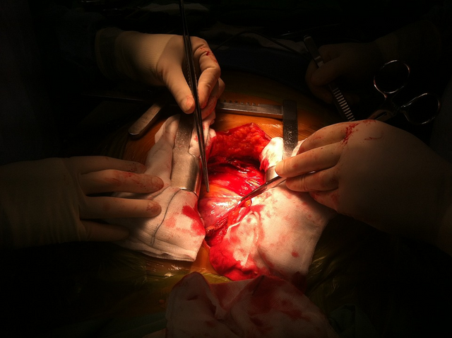
Open heart surgery. Public domain image. Source. Pixabay
The medical technique used is surgical resection, with cardiopulmonary bypass, with satisfactory results. Myxomas recur or recur more frequently in those cases that have a family history of cardiac tumors in 20% of reported cases and only 2% of individuals with sporadic presentation, ie without a family history, such recurrence after resection also usually to associate with those multifocal tumors and when the extraction thereof is incomplete from the tumor.
This type of tumor lesion, Myxoma are of good prognosis, and the survival rate as long as it is detected and treated early.
Sources of support in the publication
The Mediastinum and Its 3 Main Regions By Lynne Eldridge, MD.
Anatomy and Function of the Heart Valves
How Does the Blood Flow Through Your Heart
Several diagnoses in a patient with left atrial myxoma: Apropos of a case
Clinical presentation and treatment of cardiac myxoma in 153 patients
HARRINSON Principles of Internal Medicine, edition 16. Dennis L. Kasper et al. Part VIII. Chapter 223.

steemSTEM is a project of the chain of blocks that supports the scientific content in different areas of science. If you want to know more about this wonderful project you can join the server in discord
This article will be published at https://www.steemstem.io/

I hope you enjoyed my content.
Hey very nice article,
a few weeks ago I was thinking about cancer in the heart because I never heard about it. Now I know it definitely exists. What do you think is the reason for the low incident? You mentioned genetic reasons. What else could cause this?
Thanks in advance
Regards
Chapper
Hello ... thanks for taking the time to read my content. Actually this type of pathology is not as frequent as other pathologies, a clear example is the case of lung or breast cancer, worldwide campaigns are carried out in favor of prevention and early diagnosis.
Although it is true, until now cardiac tumors such as atrial myxoma, is a benign entity, which has an exclusive genetic-hereditary base and which some authors describe as autosomal dominant, associated with an alteration of the PRKAR1A gene, is not associated with another cause specific, unlike other pathologies, for example, lung cancer that is related to both hereditary factors and the abuse of cigarette smoking.
Greetings again thank you ..! @chappertron
I was aware of cardiac tumors. A friend on mine had one. However, I didn't know before this post that there is a primary lymphoma in heart. I looked it up, it's extremely rare, nevertheless, interesting.
Thanks for the great post.
Greetings @scienceblocks, thanks for your support and read the publication, these pathological entities are very rare, but they exist. The topic of lymphoma is very extensive. In fact, cardiac and ocular primary lymphoma have been described, very infrequent variables.
In particular, the case described in this publication is based on an atrial myxoma, which we can see in detail in the video, this being a large one, which was unleashing our clinical patient of heart failure and superior vena cava syndrome.
Thanks again for your support, success.
This post has been voted on by the SteemSTEM curation team and voting trail. It is elligible for support from @curie and @utopian-io.
If you appreciate the work we are doing, then consider supporting our witness stem.witness. Additional witness support to the curie witness and utopian-io witness would be appreciated as well.
For additional information please join us on the SteemSTEM discord and to get to know the rest of the community!
Thanks for having added @steemstem as a beneficiary to your post. This granted you a stronger support from SteemSTEM.
Thanks for having used the steemstem.io app. You got a stronger support!
Greetings, thanks for the support.
Hi @anaestrada12!
Your post was upvoted by Utopian.io in cooperation with @steemstem - supporting knowledge, innovation and technological advancement on the Steem Blockchain.
Contribute to Open Source with utopian.io
Learn how to contribute on our website and join the new open source economy.
Want to chat? Join the Utopian Community on Discord https://discord.gg/h52nFrV
Greetings, thanks for the support.
Very informative article, thanks for sharing this! I had read a bit about heart tumors before, but I admit I knew next to nothing about them, apart from the fact that they existed. I'll make sure to consider them as a possible diagnosis in cases of apparent endocarditis, despite their rarity, one never knows when that may save a life.
Posted using Partiko Android
Greetings @ mike961, in medicine not always one plus one equals two, hehehe. Actually when assessing a patient, the range of differential diagnoses must be broad and discarded one by one.
Thanks for reading the successful publication.