What You Need To Know About The Silent Thief Of Sight
Hi everyone,
Have you ever heard about glaucoma? Yup, I'm quite straight-forward, there is no point in hiding the topic of my article until the end, isn't it? So, do you know why glaucoma has the reputation of being called as the silent thief of sight? Some patients who have this kind of ailment are asymptomatic; they didn't manifest any kind of symptoms which might indicate they have glaucoma. What do you think is the outcome of this disease? Well, if patients don't seek medical treatment for glaucoma, they can end up blind. Glaucoma has been the leading cause of irreversible blindness, worldwide and it is one of the most common causes of chronic vision loss in patients (the other causes would be cataract and diabetic retinopathy).
In 2006, it was estimated that 70 million people have glaucoma whether they were formally diagnosed, suspected medically or could not care less even with all of the symptoms. It's quite difficult to get the actual number as only 10-50% of people were diagnosed while the rest appear to be asymptomatic. Out of the 70 million people, approximately 10% of that population were diagnosed with bilateral complete visual losses. Let me tell you something, the figure was quite significant and the type of visual loss inflicted on the individual with glaucoma is irreversible (permanent). If you have glaucoma, the only objective of treating it is to preserve vision, not to restore the vision which has been lost; at least for now.
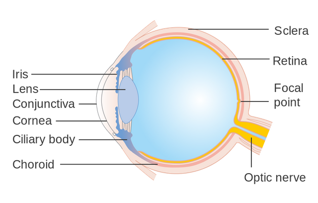
The eye might be a small organ but they are made up of various structures which would enable us to see. A structure which is called the retina, would converge and converts incoming lights into an electrical signal to be transmitted to the brain and any defects which hinder this particular capability can cause visual loss. In glaucoma, we are particularly interested in the patient's intraocular pressure. Even though there are a few glaucoma patients who are having a normal intraocular pressure, it is still one of the most common parameters used to diagnose glaucoma and monitor a patient's response towards glaucoma-related treatment.
There are two types of glaucoma:
- Open-angle glaucoma
- Closed-angle glaucoma
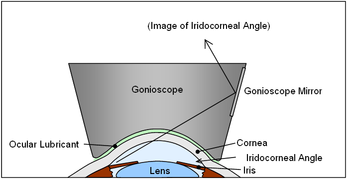
The terms "open-angle" and "closed-angle" with the glaucoma diagnosis concern with the iridocorneal angle, an angle which is formed by the convergence of iris and cornea. So what's so special about this particular spot? First of all, we need to understand the mechanism of aqueous humour clearance in which, if the clearance system is defective, can lead to glaucoma. Aqueous humour is a transparent liquid produced by a structure called the ciliary body located in the anterior segment of the eye.
This low protein liquid will assist in holding the lens in its place by occupying anterior and posterior spaces of the eyes. As it is produced continuously by the ciliary epithelium, the volume of the aqueous humour in the eye will increase (followed by the intraocular pressure). Like any fluid which occupies a certain organ, aqueous humour needs to be drained back into the circulation to maintain its volume and the intraocular pressure. It will first fill the posterior chamber, passes through the opening (which we sometimes called the aperture) called pupil into the anterior chamber and subsequently drained by 3 structures (located at the iridocorneal angle) back into the circulation:
- Trabecular meshwork (primarily)
- Schlemm's canal
- Conventional pathway through the episcleral veins
Glaucoma will ensue if there is some defect to any of the draining mechanism (usually the trabecular meshwork) which would raise the intraocular pressure. People with small eyes and shallow anterior chamber are prone to get closed-angle glaucoma. When the anterior chamber is quite shallow, the trabecular meshwork located at the iridocorneal angle was easily blocked especially by pupil dilatation. This will cause some sort of resistance to the aqueous humour outflow which would lead to an increased in intraocular pressure.
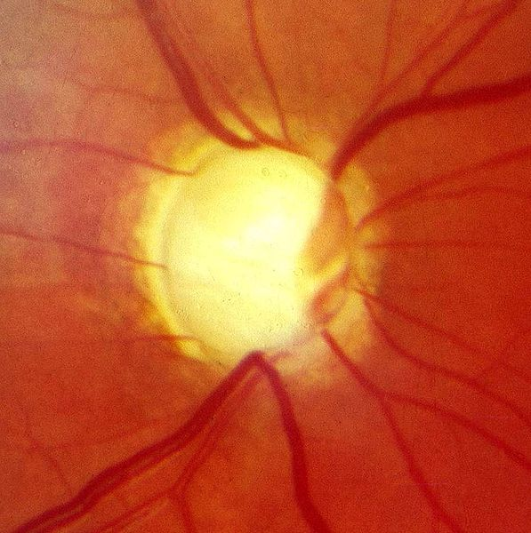
An increase in the intraocular pressure can damage the posterior structure of the eyes particularly the optic nerve head which is considered as an important structure to be monitored in glaucoma patients. The optic nerve head or optic disc consists of two significant portions which are:
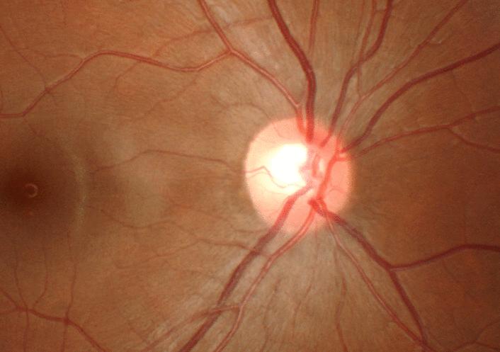
- The pale centre (optic cup)
- The slightly red kinda orangy surrounding the optic cup which is called as the neuroretinal rim.
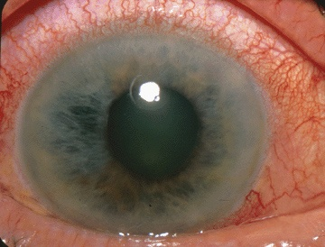
The optic disc is actually a point of exit for axons, converging into a structure known as the optic nerve. It is also filled with blood vessels which are sensitive to pressure gradient inside the eye. An increased intraocular pressure can lead to a reduced (or none) blood supply to the optic nerve head making the nerves fibre die. This is represented as an increase in diameter of the pale centre of the optic disc. For a normal people, the cup (the diameter of the pale centre) to disc (the diameter of the whole structure) ratio were between 0.3-0.5 but in glaucoma, it can be as high as 0.9.
There are of course other signs that can be observed in an optic disc of a patient with glaucoma such as bayoneting of the vessels, nasalisation of the vessels etc. but I wouldn't go so far as to confuse you with all of the details. Closed-angle glaucoma is an acute condition which is caused by an abrupt increase in the intraocular pressure. As opposed to an open angle glaucoma, they are painful and usually treated as a medical emergency.
In an open angle glaucoma, the iridocorneal angle is normal. The resistance to the outflow of the aqueous humour is caused by three factors:
- Trabecular lamellae pore size reduction
- Reduced trabecular lamellae count
- Increased extracellular materials within the network of trabecular lamellae
The outcome is the same with the closed angle glaucoma; it will cause an increase in the intraocular pressure causing damage to the axons forming the optic nerve, affecting vision. The process is painless and slowly progressing which why, up to 50% of the glaucoma population don't even realise they were having glaucoma which can lead to an irreversible nerve damage, consequently, vision loss.
Clinical presentation, challenges and treatment

Let's are honest, patients aren't textbook. That's why research is an important aspect of the medical field as there are so many contradictory cases which present with different symptoms for a particular case. Glaucoma, for example, has been known to strongly correlated with an increased in the intraocular pressure. It's textbook; when the pressure increases, the axons will be damaged and vision should be lost. However, there are many cases which have been documented in various journal platform which found that 50% of individuals with glaucoma has an intraocular pressure within the normal parameter. Some patients who have been recorded as having a high intraocular pressure for a specific duration of time were never diagnosed or found to have signs and symptoms which is associated with glaucoma. This is particularly disturbing.
There are 3 parameters which would be carried out on an individual to check whether or not, he/she has glaucoma:
- Intraocular pressure
- Visual field
- Evaluation of the optic disc
Making an early diagnosis can improve the patient's quality of life but as you can see, most of the people who are having glaucoma, don't even realised they had them. Unless they did a regular check-up at the local clinic or hospital, they won't be diagnosed until the damage has become severe and their vision has became compromised. Just like what I have stated earlier when a vision has been lost due to glaucoma, it can't be restored. The only thing that we can do is to try and preserve the rest of the functioning vision.
The pattern of visual loss was usually from the periphery to the central visual field. In theory, to provide an ideal care for a patient would be to identify certain risk factors which could lead to the development of glaucoma but in reality, when a patient is diagnosed with glaucoma, as much as 50% of the retinal ganglion cells have been lost, courtesy of the increased in the intraocular pressure. Medical doctors depend heavily on the chief complaint of a patient to make an accurate diagnosis. Glaucoma is a progressive disease which can potentially cause visual damage in a matter of years, and as the process was pain-free (open-angle glaucoma), there is nothing to complain about, until the damage has become severe. Making an early diagnosis is quite an arduous task and by the time, they were diagnosed, a significant portion of vision has been stolen from the patient.
The assessment of optic discs was done by an ophthalmoscope. It is a useful device to assess the integrity of the optic disc of a patient but the conclusion might differ from one medical doctor to another; the assessment is quite subjective and will require years of training. The role played by a physician is important. People who have risk factors i.e. high intraocular pressure, symptoms related to glaucoma or family history of glaucoma should be referred to an ophthalmologist for further assessments.
There are three groups of treatment modalities:
- Medication
- Laser trabeculoplasty
- Surgical trabeculectomy
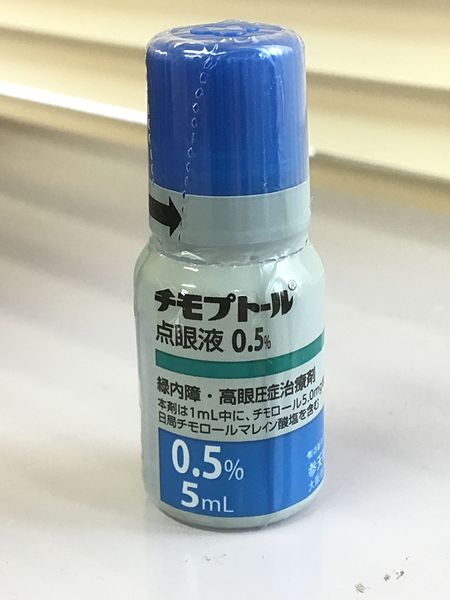
When I was in the medical school, we have been thought to answer questions pertaining to the management in a series of organised steps. If it is a question related to the investigation, the step should be biochemical, imaging and special test and if the question related to management, the first step would be to prescribe medications, keeping surgical intervention as the last resort. In glaucoma, the aim is to maintain a low intraocular pressure to minimise damage to the optic discs. There are a few types of medications which are being used to treat glaucoma which are:
- Beta blockers: To reduce the aqueous humour secretion
- Parasympathomimetic: To increase the outflow of aqueous humour
- Carbonic anhydrase: To reduce the aqueous humour secretion
- Prostaglandins analogue: To increase the outflow of aqueous humour
The medication can be used in combination with each other to augment the action of that particular drug but if the intraocular pressure remains high, the alternatives would be to perform laser trabeculoplasty or proceed with trabeculectomy. Laser surgery will provide symptomatic relieved to the intraocular pressure which would prevent the symptoms of glaucoma from worsening. Some studies have shown this laser treatment to be effective in treating an early onset glaucoma, but not the late onset. It is usually performed to treat a close angle glaucoma, providing an immediate pain relief due to an increase in the intraocular pressure as a result of trabecular meshwork blockage at the iridocorneal angle.
If none of the above treatments works, a fistula can be created between the subconjunctival space and the anterior chamber, draining the aqueous humour which subsequently will reduce the intraocular pressure in a procedure known as the trabeculectomy. As you can see, all of the treatment modalities aimed at lowering the intraocular pressure and not restoring the vision. There is no treatment available for optic nerve ischaemia just yet. In some patients, despite successful effort at lowering their intraocular pressure, their vision got worse day by day. Once again, a patient is not a textbook. It is not even an exam question that I have took in medical school to get a medical degree. The future of medicine will depend on research and let's hope someday, we can actually restore the integrity of the optic nerve head providing people with the opportunity to see again.
Source
- Optic Nerve Cupping
- The Pathophysiology and Treatment of Glaucoma
- Acute primary angle-closure: long-term intraocular pressure outcome in Asian eyes.
- Prevalence of open-angle glaucoma among adults in the United States.


SteemSTEM is a community project with the goal to promote and support Science, Technology, Engineering and Mathematics on the Steem blockchain. If you wish to support the steemSTEM project you can:
Contribute STEM content using the #steemstem tag | Support steemstem authors | Join our curation trail | Visit our Discord community | Delegate SP to steemstem
You really know how to tickle my eye procedure sensibilities :D
Took a while to read but, as usually, your article is good. It's too bad that I am not a medical doctor or have any real experience with procedures, but I can trust your expertise :P
Ocular health is important as it's probably the most relied on sense. So, for anyone reading this, don't forget to schedule a check from time to time, especially with age. Always wear protective eyewear when exposed to sun, and always protect your eyes when working with dust, chemicals and other sort of contaminants.
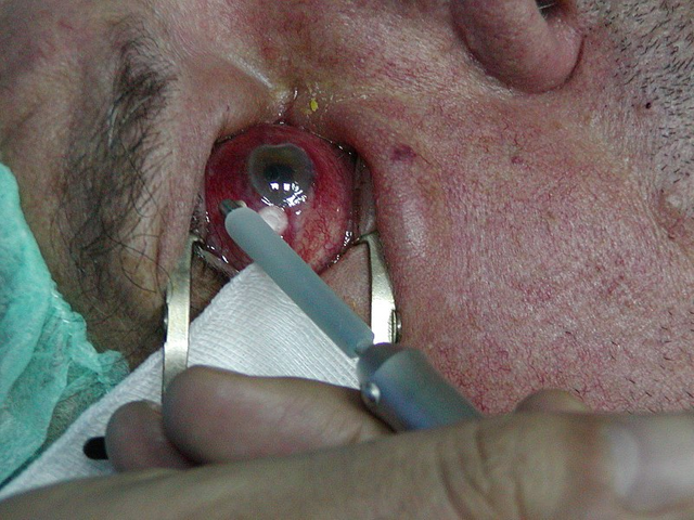
For some reason, I hate pictures with eyes... brrr.... I am happy the ugliest picture came at the end of the post (otherwise, I would have not reads it :p ). If I would not have any visceral problem with eyes, I would have loved to study the physics behind that :)
@lemouth: Hahaha. Yeah, even though I have dealt with hundreds of eyes throughout my clinical practice, I'm still uncomfortable with eyes surgery. I guess ophthalmology is not for me.
@alexdory: Yeah, you're right. Eyes might be small but I can't stress enough the importance of it. Losing sight would affect the quality of life of a patient greatly.
Lucky I have chosen another field of science :D
Sight is probably the most important of our senses, and diseases like this one must be the worst for the people who suffers them.
Either way, as you mentioned in the article, at least there are procedures that can help the patients before they have lost their vision.
The last picture is really shocking, it must be hard for the doctor to manipulate the eye of another person like that.
Cheers @conficker!
Well, it will get easier with practice. Unfortunately, we can't restore the vision which has been lost, only preserve whatever left.