MICROSCOPE; A "TREASURE" AND VITAL INSTRUMENT IN DIAGNOSTIC MEDICINE
 )
https://pixabay.com/en/scientist-drugstore-microscope-chemistry-2141259/
)
https://pixabay.com/en/scientist-drugstore-microscope-chemistry-2141259/
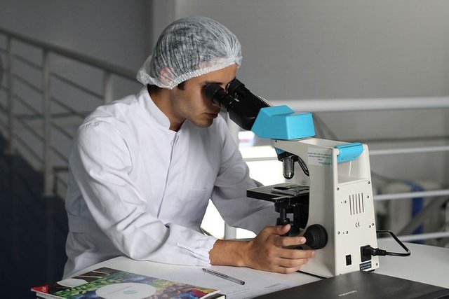
Hello steemians,its a privilege to be back in your mist and i hope to consistently contribute my little knowledge in this awesome community.In our world today in comparison with mobile phone in the technology sector, the microscope in the medical field is an equipment that has continue to evolve and improve in its services and role in modern day medicine.the uses,importance and reliability of microscope in daignostic medicine has increase tremendously,it has increase the speed,validity and reliability of results. Many times if you go to the laboratory you find scientist spending hours with the microscope in the lab carrying out routine test and most of whom are now fond of it just like the way most of us are fond of our phones. Also the role of a microscope in the laboratory is inexustible it ranges from microbiology field where it has made it possible for us to diagnose microbes affecting man to that of haematology section where it is important in carrying out routine test such as total white blood cell count etc,in histopathology also it aid in studying the intrinsic nature of tissues pathologically affected.Now permit me to disect and explore this awesome and amazing instrument to enable you appreciate more the role it play in our lilfes medically.
MICROSCOPE AND MICROSCOPY
A microscope is an instrument used to see substance or objects that are too small or tiny to be seen by the naked eye. Microscopy is therefore the science of investigating small objects and structures using a microscope. Microscopic means invisible to the eye noneless aided by a microscope. For one to understand how a microscope works or operates one need to know or understand how the lenses formed images from beams of light or electrons passing through it.also refraction,focal length and refractive index are other features of a lens that is important.
A magnifying glass is also seen as a microscope(simple microscope)because it is able to magnify or enlarge tiny objects and the more fancy microscope you know is called a compound microscope .Therefore a microscope is an instrument that uses a lens to view or produce images of tiny objects. A picture from a microscope is called a micrograph.
HISTORY
The story of the invention of microscope has been controversial because of many claims of it invention by many authors and scholars.though notably in the history of microscope development many scientist had contributed from Galileo galilie to Giovanni faber who coined the word microscope to marcelo malpighi(father of histology)and to anthonie van leeuwenhoek whose contribution is immensely appreciated in the development of the microscope.
IMPORTANT PARAMETERS IN MICROSCOPY; magnification and resolution.
MAGNIFICATION:this is a measure of how large or big a microscope is able to make a tiny object appear.it simply involves how much enlargement can be done by a microscope on an image.when an image is said to have a magnification of ×400 it mean the initial image of an object has been increase 400 times(i.e 1cm is now 400cm).
RESOLUTION:this involves the smallest distance between two objects by which both can be separeted and still exist as separate entities.it is a measure of how clear and detail your image is.most times objects or specimens view with the microscope are tiny and usually appear blur when they are close or clusters,but with the resolving power of the microscope can be made clear.
These two parameters are importants and go handy,one cant go without the other for a good and nice image to be produce.A magnify can be blur and a clear image can be too tiny.
TYPES OF MICROSCOPES
microscopes are classify based on different features;1.number of eyepiece (could be monocular or binocular) 2.source of beams wavelength(light or electrons).
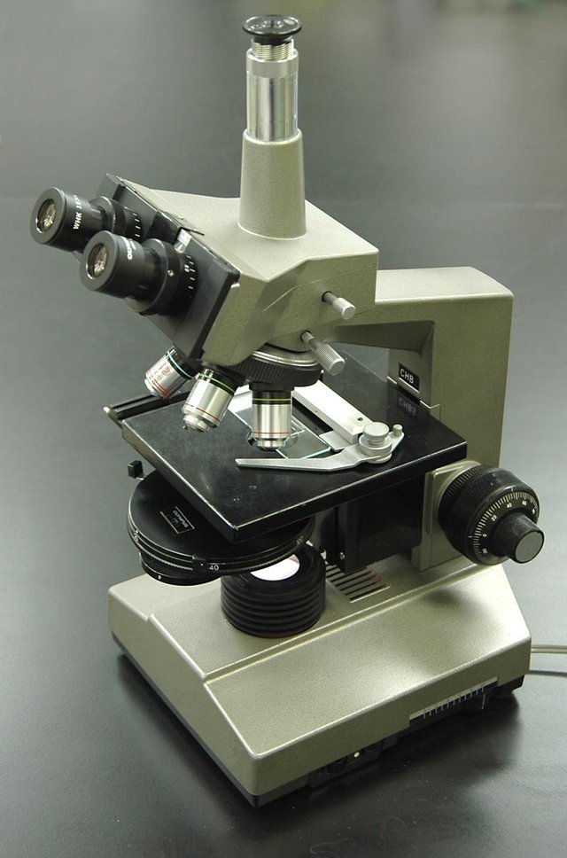
LIGHT OR OPTICAL MICROSCOPES
These are microscopes that make use of visible light to magnify objects,the visible light pass through the specimen and lens system and then produce images in the eyes of the observer,photographic plate or captured digitally.most student laboratory microscope are usually that of optical microscope.their are varieties of light microscope which are bright-field microscope,dark-field microscope,phase-contrast microscope and fluorescence microscope e.t.c.
Bright-field microscope:most light microscopes we see are bright-field microscopes which form dark images on a bright surrounding or background.most specimen view by this type of microscope must be stained fixed or live organisms.usually a light source or mirror transmit lights through the view specimen,making the stained specimen dark and its surrounding bright.also a condenser which regulate the light passing through the specimen is also seen beneath the stage that hold the specimen.
Dark-field microscopes: this type of light microscope only produce image from light refracted or reflected by the specimen and is used to view unstained,living cells and organisms.it involve the use of dark-field stop underneath the condenser. A bright image is formed on a dark background.Dark-field microscope is usually use in identifying treponema pallidum

Phase-contrast microscopes: it works on the principle of slight differences in refractive index and cell density and converts this variations in light intensity into detectable images,use to view living cells which are not stained and are difficult observing with a bright-field microscope.this kind of microscope make use of a condenser that allow light to pass the specimen only.
Fluorescence microscope: some microbes or molecules are autofluorescent(being able to emit light when excited) in nature and thats were this type of microscope comes in. An objects or microorganism can be view clearly by this microscope when its has absorb energy from a light source e.g ultraviolet,blue or violet rays and as such become excited to emits light and a dark-field condenser is use so that only the light emitted from the organism or objects will be able to form an image in the lenses.this object or organism that are being viewed maybe stained with a dye called fluorochrome when they are not autofluorescent in nature.most migrograph produce from this type of microscope are usually very beautiful and clear cause these objects or oganisms emits different colours be it blue,green or red e.t.c.
ELECTRON MICROSCOPES
For many years scientists continued to use the light microscopes despite its limitations which majorly is its disability to resolute less than 0.2um which is the highest resolution the best light microscope can offer.as a result scientists could view the morphological and external structural features of a bacteria which is about 1um but couldnot view its internal structures i.e its organelles.
Now with the advent of electron microscope this limitation has been overcome. An electron microscope is a type of microscope which make use of electron beams instead of light to produce images of microorganisms.it has a better resolution than light microscopes because the wavelength of beams produce by electron are shorter than light beams.
Transmission electron microscope and scanning electron microscope are the types of electron microdcopes.
Transmission microscopes: these microscopes works by transmitting or passing electrons through the specimen that is being viewed.it works in a similar technique as light except that it uses a heated tungsten filaments in an electron gun to produce electron beams instead of light source,it uses a magnetic lens instead of a glass lens cause electrons cant pass through glass and also the image is formed on a fluorescent screen.It is important to note also that the specimen to be observe must be prepared i.e its thickness must be reduced to 20nm-100nm and also coated with heavy metals to prevent the specimen from absorbing or scattering the electrons.a condenser is use to ensure that the electrons pass through the specimen.
Scanning electron microscopes: Instead of electrons passing through the specimen this type of electron microscope work by forming images from electrons emitted from the surfaces of specimen after it has been scanned by electrons produce from the electron gun.with the use of a detector,scintillator and a photomultiplier it produce an image from a cathode-ray tube.
CONCLUSION
Microscope is an invaluable tool in modern day daignostic medcine, imagine a world without the use of microscope then you will appreciate more its impacts in our our society today. thanks to microscope the field of forensic science has help solve crime issues and other critical problems in our world today.thanks to microscope we able daignose diseases conditions of our love ones.with microscope science has become easier and fun.
thanks for reading until next time!
Thank you for your contribution. Dont forget to link references and sources when applicable!
=======================================================================================
This post was upvoted and resteemed by @Steemgridcoin with the aim of promoting discussions surrounding Gridcoin and science.
This service is free. Please follow @steemgridcoin if you want to support this initiative.
Have a nice day. :)
First of all, if you think a microscope is kinda important in medical diagnostic processes, then you are right. It is, but I think the definition you provided for the term microscopy needs to be modified a little. I would say, it is not to investigate small things but rather trying to see things which can't be perceived by the naked eyes. Of course, previously we can describe it as a technique to view all kind of microorganism but in 1999, scientists have discovered a bacterium which is quite large and can be seen without using a microscope.
I mean, if the object is small but still can be seen by our naked eyes, we wouldn't need a microscope, do we?
thanks for this wonderful contribution ...you are indeed right.
Recent related articles
Older related articles
Interesteem(@interesteem) is a service that recommends related articles using DeepLearning.
Please write an article with #interesteem tag.
Congratulations @makovicloyal! You have completed the following achievement on the Steem blockchain and have been rewarded with new badge(s) :
Click on the badge to view your Board of Honor.
If you no longer want to receive notifications, reply to this comment with the word
STOPCongratulations @makovicloyal! You have completed the following achievement on the Steem blockchain and have been rewarded with new badge(s) :
Click on the badge to view your Board of Honor.
If you no longer want to receive notifications, reply to this comment with the word
STOP