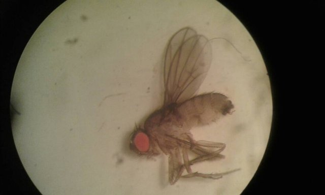
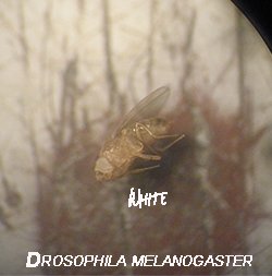
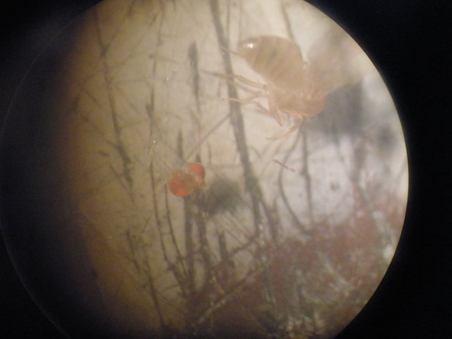
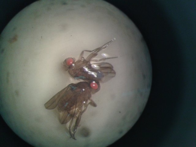
EYE PIGMENTS CHROMATOGRAPHY in Drosophila Melanogaster- Fruit fly.
This particular species of fly was taken because of its characteristics, since it has a low number of chromosomes, easily recognizable phenotypic variability and whose chromosomal location is known, as well as easy growth in simple culture media, which facilitated the experiment. and determine the pigments responsible for the coloration of eyes and body from a thin layer chromatography.
Introduction.
Theories of inheritance date back to the fourth and fifth centuries BC. Genetics (the scientific study of inheritance) did not really begin until the eighteenth and nineteenth centuries. Basically the transmission from generation to generation of certain characters in plants and animals was observed in order to provide a rational basis for improving crops and livestock.
The most significant advance in the field of genetics was provided by the Austrian monk Gregor Johann Mendel (1822-1884), who observed certain specific characteristics in pea plants, counting the number of individuals in which these traits appeared throughout of several generations. Concentrating on just a few characters and determining in what proportion of each generation they were present, he was able to demonstrate the basic specific patterns of inheritance. The discrete character and independent segregation of the genetic characteristics that he observed are now known as the "Laws of Mendel's inheritance" and apply to most genetic systems.
One of the most significant contributions to genetics was made by Thomas Hunt Morgan (1866-1945), who recognized the presence of sex chromosomes and what is known in genetics as "sex-linked inheritance". He also showed that Mendelian factors (genes) were arranged linearly on the chromosomes. The experiments conducted by Morgan and his colleagues also revealed the genetic basis of sex determination. Morgan continued his experiments and demonstrated in his "Theory of Genes" that the genes are united in different groups of chaining, and that the alleles (pairs of genes that affect the same character) are exchanged or cross-linked within the same group.
Experimental procedure.
Chromatography of pigments of eyes and bodies of Drosophila speck.
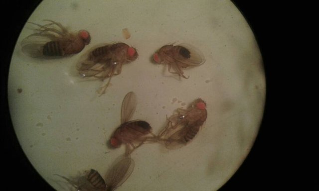
We take 4 filter papers of 23 cm wide by 28 cm high, in the line of application we sow the heads and the bodies each one in different points, as it is described next with the symbols used in the laboratory guide to name the mutations:
Paper # 1. Fly heads: + ♀ w♀ ♀we ♀wa ♀wa / w ♀ we / wa.
Paper # 2. Bodies of fly heads placed on paper # 1.
Paper # 3. Fly heads: + ♀ w♀ ♂w ♀Cu ♀bw ♀Cu / bw.
Paper # 4: body of the fly heads placed on paper # 3.
After the planting we placed the papers in the buckets containing the solvent system 20: 3: 7 butyl alcohol / glacial cetico acid / distilled water. Once the chromatography run was developed, the papers were visualized, using a UV light viewer.
CHROMATOGRAPHY OF EYE PIGMENTS:
In specimens of wild D.melanogaster, the pigmentation of the eyes is due to the presence of two types of pigments: pteridins and ommochromes; the former are brightly colored compounds that fluoresce and emit a characteristic color light when viewed under UV light. The ommochromes are brown and do not fluoresce in UV light. The production of each type of pigment involves a sequence of known biosynthetic steps. The precursor of the pteridines is 2-amino-4-hydroxypteridine, while that of ommochromes is the amino acid tryptophan. Once formed these pigments must be fixed on the protein granules of the ommatidia that make up the facets of the eyes. The set of these pigments in wild individuals makes the eyes are red. In the fixation of the pigments, the activity of another gene independent of those genes related to biosynthesis intervenes. It is the wild allele "white", the activity of the gene involved in the process of fixing the pigments to the granules of ommatidium protein. Thus, the w / w mutant females and the hemigo w / Y males are unable to fix ocular pigments, and although they can biosynthesize them, their eyes are white. This result is reflected in the chromatograms performed. A mutant strain in a determined metabolic sequence, as is the case of the formation of pigments, can be recognized because it lacks the final product of the biosynthetic sequence, however, it presents the compound that precedes the mutated biosynthetic step in the sequence.By chromatography on paper and using a system of suitable organic solvents, it was possible to separate the pigments into different identifiable spots.
For a wild fly strain chromatography allows the separation of 7 different pteridines: drosopterin, golden yellow; isoxantopterin, purple; sepiapterin, lemon yellow; 2-amino-4-hydroxypterin, blue in color; biopterin, violet blue; xantopterina, blue-green color and isosepiapterina, orange yellow. The eye-color mutants of D. melanogaster present a pteridin pattern different from the pattern of the wild strain. To discern phenomena of allelic interaction, it is very important to establish the level of observation of the individuals under study, and thus be able to establish the relationship of dominance between a gene and its allele. In the case of homologous individuals (AA or aa), the joint expression of both identical alleles leads to the expression of a characteristic, dominant or recessive phenotype. The heterozygous Aa individuals may be phenotypically identical to the dominant AA homociples, however, when the phenotypic analysis is performed at another level of observation, for example at the biochemical level, the phenotype differences between individuals AA and Aa can be established. . The level of observation of a heterozygous individual may reveal the activity or absence of activity of a recessive gene. An analysis of the products synthesized by the genes in question, will allow to recognize the activity or hidden inactivity of the recessive allelic gene that is masked by the presence of the dominant gene product in the heterozygote, this will allow to establish differences between homologous and heterozygous individuals.
Results and discussions.
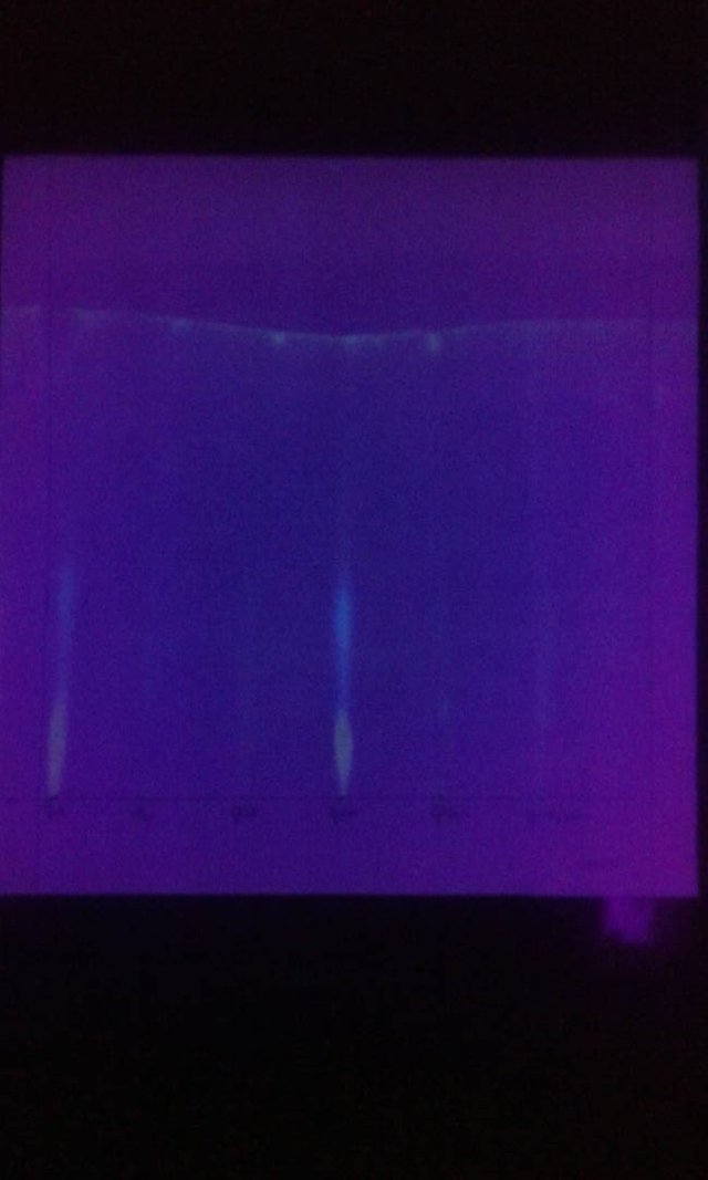
In specimens of wild D. melanogaster, the pigmentation of the eyes is due to the presence of two types of pigments: pteridins and ommochromes; the former are brightly colored compounds that fluoresce and emit a characteristic color light when viewed under UV light. The ommochromes are brown and do not fluoresce in UV light. The production of each type of pigment involves a sequence of known biosynthetic steps. The precursor of the pteridines is 2-amino-4-hydroxypteridine, while that of ommochromes is the amino acid tryptophan. Once formed these pigments must be fixed on the protein granules of the ommatidia that make up the facets of the eyes. The set of these pigments in wild individuals makes the eyes are red. In the fixation of the pigments, the activity of another gene independent of those genes related to biosynthesis intervenes. It is the wild allele "white", the activity of the gene involved in the process of fixing the pigments to the granules of ommatidium protein. Thus, the w / w mutant females and the hemigo w / Y males are unable to fix ocular pigments, and although they can biosynthesize them, their eyes are white. This result is reflected in the chromatograms performed (figure 5). A mutant strain in a determined metabolic sequence, as is the case of the formation of pigments, can be recognized because it lacks the final product of the biosynthetic sequence, however, it presents the compound that precedes the mutated biosynthetic step in the sequence.
By chromatography on paper and using a system of suitable organic solvents, it was possible to separate the pigments into different identifiable spots. For a strain of wild flies, chromatography allows the separation of 7 different pteridines: drosopterin, golden yellow; isoxantopterin, purple; sepiapterin, lemon yellow; 2-amino-4-hydroxypterin, blue in color; biopterin, violet blue; xantopterina, blue-green color and isosepiapterina, orange yellow. The eye-color mutants of D. melanogaster present a pteridin pattern different from the pattern of the wild strain. To discern phenomena of allelic interaction, it is very important to establish the level of observation of the individuals under study, and thus be able to establish the relationship of dominance between a gene and its allele. In the case of homologous individuals (AA or aa), the joint expression of both identical alleles leads to the expression of a characteristic, dominant or recessive phenotype. The heterozygous Aa individuals may be phenotypically identical to the dominant AA homociples, however, when the phenotypic analysis is performed at another level of observation, for example at the biochemical level, the phenotype differences between individuals AA and Aa can be established. . The level of observation of a heterozygous individual may reveal the activity or absence of activity of a recessive gene. An analysis of the products synthesized by the genes in question, will allow to recognize the activity or hidden inactivity of the recessive allelic gene that is masked by the presence of the product of the dominant gene in the heterozygote, this will allow to establish differences between homologous and heterozygous individuals.
Conclusions.
It was possible to obtain a chromatography of the pteridin pattern of normal and mutant strains of D. melanogaster.
Differences in the pteridin patterns of the different mutant strains were evidenced. There were also variations between sexes.
Differences were established at the biochemical level between homologous and heterozygous individuals.
Annexes.
References.
• Anthony Griffiths 2008. Genetics. Interamerican McGraw-Hill of Spain. S.A.U. 9th edition. Madrid. Spain.
• William Klug, 2013. Concepts of Genetics. Pearson Education, S.A. 10th edition. Madrid.
• Daniel, Wayne W. 2009 Biostatistics. Editorial Limusa, S.A. 4th edition. Mexico.
• Genetics teaching unit. Guide of the General Genetics Laboratory of the Department of Cell Biology. Central Unit of Venezuela.
I hope you liked this publication, thank you for visiting my post.
Work done at the Central University of Venezuela by @Norihany. The photographs are taken with a samsung s4 phone by @Norihany.
Somewhere in the world.
Chic article. I learned a lot of interesting and cognitive. I'm screwed up with you, I'll be glad to reciprocal subscription))
muy bueno tu trabajo de hoy nori
Gracias :))Congratulations! This post has been upvoted from the communal account, @minnowsupport, by Norihany from the Minnow Support Project. It's a witness project run by aggroed, ausbitbank, teamsteem, theprophet0, someguy123, neoxian, followbtcnews, and netuoso. The goal is to help Steemit grow by supporting Minnows. Please find us at the Peace, Abundance, and Liberty Network (PALnet) Discord Channel. It's a completely public and open space to all members of the Steemit community who voluntarily choose to be there.
If you would like to delegate to the Minnow Support Project you can do so by clicking on the following links: 50SP, 100SP, 250SP, 500SP, 1000SP, 5000SP.
Be sure to leave at least 50SP undelegated on your account.