Vision: Who Said We See With Our Eyes?
Introduction
Vision(sense of sight), one of the special sensory modalities in the body and the most important of them all if I may award, is brought about by the working together of different things which, for some reason whatsoever are seen to be concentrated in the head. This does not in any sense mean that we see with our heads. Or, does it?
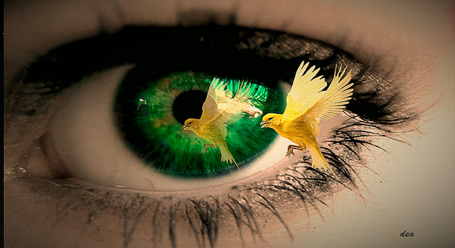
The Eye Focusing on A Flying Bird(License: CC-BY 2.0, Author: Claudia Dea]:
Flickr
Why then does it seem so convenient and comfortable to say that “we see with our eyes” when the eye only houses about 70% of the total media through which vision is made possible and not the whole 100; when it also behaves like every other receptor in the body, generating specific signals(receptor potential) and sending them to the central nervous system(in this case, the brain) for interpretation and response; when it acts only as a means to an end by having to transfer its information to a final destination (the cerebral cortex) before vision can be accomplished and not capture the image, process it, save it, and possibly give a response to the body?
Well, in this post, I will be dealing with how vision is made possible and the components that work together to accomplish this awesome act of life so we really can see the total role of the eye in the whole process and get to be sure if truly, we see with our eyes.
How Really Does The Eye Refine an image?
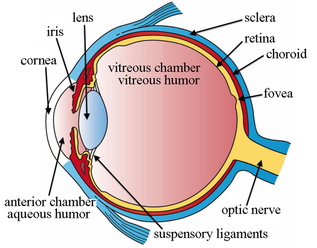 The Eye, Showing its Diferent Components (License: CC-BY 3.0, Author: Holly Fischer]:
Wikipedia Commons
The Eye, Showing its Diferent Components (License: CC-BY 3.0, Author: Holly Fischer]:
Wikipedia Commons 
The eye, anatomically known as the ocular apparatus, is somewhat spherical in shape and is made up of an outer coat (sclera) which offers protection to the content of the eye and holds the eyeball in shape, a middle layer (choroid) which maintains and nourishes the eye and an inner layer (retina) adapted to receive images and generate impulse to be sent to the cerebral cortex.
when the eye is focused on an object, light rays falling on the object are reflected by the object into the eye. As a matter of fact, the first part of the eye that the rays come in contact with is the cornea
The cornea is the outermost coat of the eye which is a continuation of the sclera in front of the eye. It is enhanced with transparency to allow the easy passage of light and is surprisingly supplied with oxygen directly from the atmospheric air
Once the light rays have successfully crossed the cornea, they pass through an aperture called the pupil which regulates the amount of light entering the eye by either constricting to reduce the amount or dilating to increase it. This particular function of the pupil plays a major role in the adaptation of the eye when light is extremely bright or when it is dark.
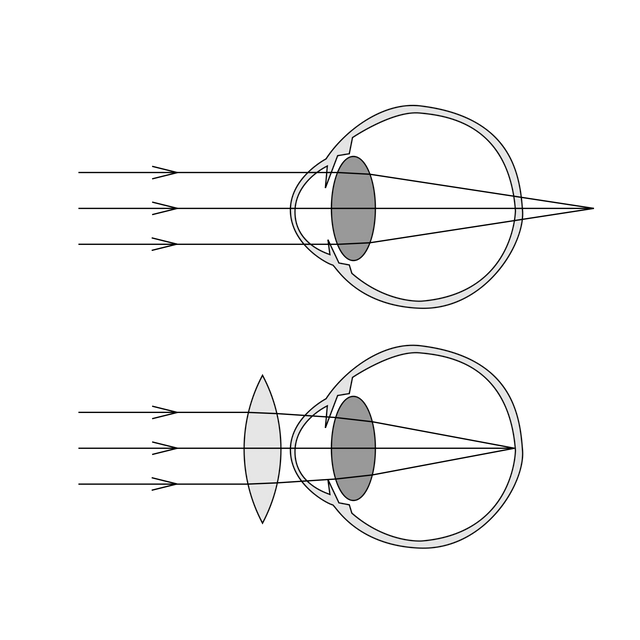 A diagramatic representation of long sightedness (License: CC-BY-SA 1.0, Author: CryptWizard]:
Wikipedia Commons
A diagramatic representation of long sightedness (License: CC-BY-SA 1.0, Author: CryptWizard]:
Wikipedia Commons 
At this point, the lens presents itself as the next gateway. It is one of the most important components of the eye as it provides the highest amount of refraction (compared to the cornea, aqueous humour or vitreous humour) to the light rays so they can converge perfectly on the retina. Failure of the rays to converge on the retina as they are supposed to is what brings about long sightedness and short sightedness
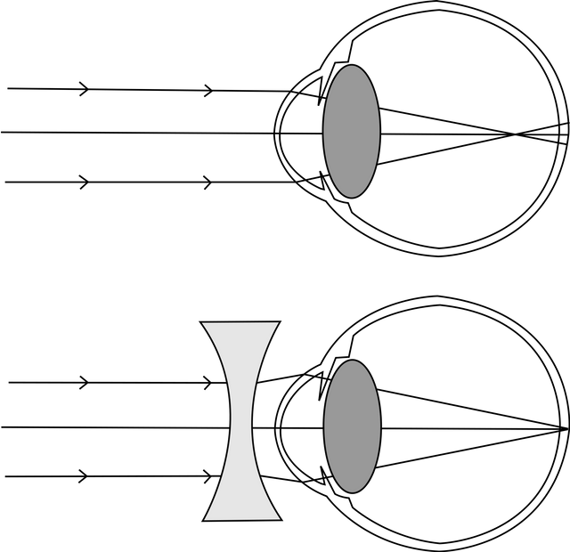 A diagramatic representation of short sightedness (License: CC-BY-SA 1.0, Author: CryptWizard]:
Wikipedia Commons
A diagramatic representation of short sightedness (License: CC-BY-SA 1.0, Author: CryptWizard]:
Wikipedia Commons 
long sightedness (hypermetropia) is a condition where an individual can only see far objects, as the light rays from a near object are made to focus at a point behind the retina while short sightedness (myopia) is a condition where an individual can only see near objects as the rays of light from a far object are made to fall in front of the retina. Myopia arises as a result of too much refraction while hypermetropia is as a result of not enough refraction
Right about now, the rays pass through the vitreous humour to reach the retina.
For a clear and elaborate understanding on how the retina accomplishes the task of transferring the light rays into electrical impulse, let us take a walk through the retina itself.
The retina is made up primarily of ten layers. These layers have different functions and house different cells. It would be my utmost pleasure and happiness to bore you with the details of the whole ten layers, but for the purpose of this post, i only want to bring into consideration, the pigment layer and the four processing cells of the retina which include the Rods, Cones, Bipolar cells and the Ganglion cells.
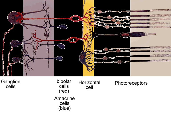
layers of the retina showing how the four phototransduction cells are interconnected (License: CC-BY-SA 3.0]:
Wikipedia Commons
The pigment layer is blessed with enormous amount of melanin and it appears black due to the presence of this pigment. The function of this layer in vision cannot be overemphasized. When light rays get to the retina, this layer absorbs the light and prevents the light rays from being reflected in all directions. This way, the light rays only excite a specific amount of rods and cones and not a large number as it would have if the pigment layer were to be absent. This is one of the reasons why albinos seem to have difficulties getting a clear image of an object. When the light rays get to the retina, they are reflected in all direction, exciting a greater number of cones and rods than they are supposed to. This causes loss of acuity and ability to get a clear image of the object.
As the light rays hit the pigment layer, the rods and cones which are in the layer just below the pigment layer, respond to the light. The rods are very sensitive to light and have a low threshold. They can be excited by light of a very low intensity, even in the dark while the cones have a very high threshold for light and can only be excited by light with intensity that has reached their threshold. The rods are responsible for black and white vision because they do not define the visual boundaries of the light rays in which they are responding to in terms of color while the cones are responsible for colored vision as they define every boundary.
Light causes a response from the receptor cells by triggering a chemical process in them which elicits a receptor potential
chemistry of vision
As a matter of fact, an image is not just put in a bag and dragged up to the brain. It is transformed to an electrical form by a chemical means just like what every other organ in the body does.
The rods and cones contain chemicals that decompose on exposure to light. The decomposition of these chemicals (known as rhodopsin in rods and color pigment in cones) causes their cell membranes to hyperpolarize instead of depolarize as it happens in other cells. This hyperpolarization causes the inhibition of the neurotransmitter glutamate which is being produced continuously in the absence of light.
This reduction in the production of glutamate causes the loss in inhibition of the bipolar cells which are connected to the photoreceptor cells. This in turn allows the bipolar cells to sense the rising and falling of light intensity and excite the ganglion cells to elicit action potential.
Once action potential has been elicited, it travels through the axons of the ganglion cells which form the optic nerve to leave the retina.
The image is finally done at the retina, is the story over now?
Of course not. It has just begun and in fact, this is exactly my point!
The retina only acts as a packager, remodeling the image and sending it to the real place where it is being perceived as the image it is.
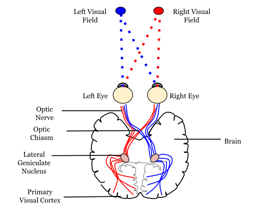 image showing the path of an image leaving the retina (License: CC-BY-SA 4.0, Author: Mads00]:
Wikipedia Commons
image showing the path of an image leaving the retina (License: CC-BY-SA 4.0, Author: Mads00]:
Wikipedia Commons 
At this point, the image is transferred by the optic nerve through the optic chiasm ( a point of crossing over between the two optic nerves of both eyes. From the point of crossing, the optic nerve now is no more known as the optic nerve but is now known as the optic tract.
This optic tract carries the image to a point in the thalamus called the lateral geniculate body. The image is the transferred from here by the optic radiation to the final destination, the occipital lobe of the cerebrum.
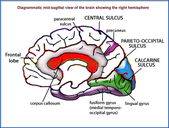 The Various Parts of the Cerbral Cortex (License: CC-BY-3.0, Author: Vaughan]:
Wikipedia Commons
The Various Parts of the Cerbral Cortex (License: CC-BY-3.0, Author: Vaughan]:
Wikipedia Commons 
On the occipital lobe of the cerebrum, there is an area known as the calcarine sulcus which is also known as the Primary visual cortex. This is the precise location where images from the retina are finally taken to and are interpreted to be seen by the individual.
This point is located at a point popularly known as the occiput (back of the head) and is responsible for the stars seen when one hits the back of the head
sumary
Vision is one of the most complex and useful processes in life. it utilizes quite a number of organs and tissues to bring about the ability to see. Though our language has found a way of overruling the main contributor in this lovely process, the fact still stands. Injury to any part of the visual path way or optic nerve will always cause an impairment in the ability to see clearly and i guess this explains it all; that what we call "seeing with our eyes", is actually "seeing with our brains"
References
All images are licensed under creative commons and eligible for commercial use. Thanks for reading
The Visual System retrieved on June 7th, 2018
Layers of the Retina retrieved on June 7th, 2018
Transformation of image to electrical impulse retrieved on June 7th, 2018
Moore, K. L., & Dalley, A. F. (1999). Clinically oriented anatomy. Philadelphia: Lippincott Williams & Wilkin
Guyton, A. C., & Hall, J. E. 1. (2006). Textbook of medical physiology (11th ed.). Philadelphia : New Delhi: Elsevier Saunders.
Moore, K. L., Agur, A. M. R., Moore, K. L., & Agur, A. M. R. (2002). Essential clinical anatomy. Philadelphia: Lippincott Williams & Wilkins
Sembulingam K., Sembulingam M.. Cardiac output, in Essentials of Medical physiology, ed 5. New Delhi; Jaypee Brothers Medical Publishers (p) ltd, 2010, p 553.
Image Sources
Hello young Steemian! You started well, with the intriguing and interesting title. But after, it was a very broad text with some basic anatomy. For STEM articles, don't be shy, show us some interesting numbers, or find some new discoveries because there is a ton of post with broad scope, every single day. Try to be original and fresh.
thanks a lot.
i appreciate the advice
Go here https://steemit.com/@a-a-a to get your post resteemed to over 72,000 followers.
detailed write up @purelyscience.
Talking about the eyes, is there a reason why we have tear drops when we'v been glued to screens for long?
Is it dangerous? so many adolescent age individuals are complaining about this
is there a solution to this??
its mainly stress.... your eyes being glued to the screen exposes the eyes to so much amount of light for a long period
using a shade or reducing brightness of screen can help reduce this stress
Hi @purelyscience!
Your post was upvoted by utopian.io in cooperation with steemstem - supporting knowledge, innovation and technological advancement on the Steem Blockchain.
Contribute to Open Source with utopian.io
Learn how to contribute on our website and join the new open source economy.
Want to chat? Join the Utopian Community on Discord https://discord.gg/h52nFrV