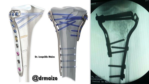
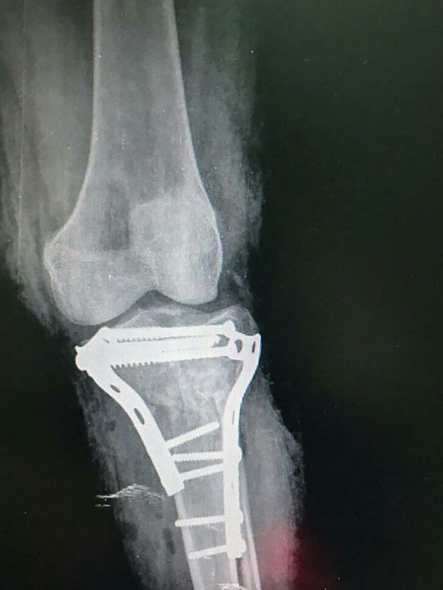
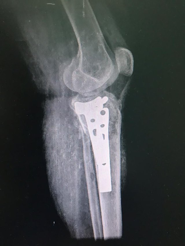
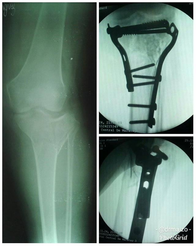
Treatment of a tibial plateau fracture. Surgery Day
The Tibial Plateau
In this post, we try to bring the reader closer to the functioning of the knee in general, and specifically the area belonging to the plateaus, through the anatomical study of it, as well as making a special interest in this injury, as well as its most effective rehabilitation techniques.
The fractures of the tibial plateaus constitute a fairly frequent traumatic injury, mainly caused by traffic accidents, which produce an increasing number of injuries every day due to their increase at the expense of the development of high speeds in motor vehicles.
There is a group of these fractures that can be resolved with conservative treatment, but in cases where there is local compression, fissure compression or complete condylea depression, the degree of compression must be taken into account, because if it is greater than 10 mm or even greater than 6 mm in young patients, most authors recommend surgery with elevation of the depressed area, filling of the resulting cavity with bone graft and restoration of the joint.

Production Mechanism
Tibial plateau fractures are caused by combined mechanisms of axial compression and valgus or varus, in turn as a result of falls from heights, traffic accidents and sports-related activities.
Fractures of the lateral tibial saucer are the most frequent according to Holh's statistics, et al from 55% to 70%, continue in frequency those of the medial saucer with a range from 10% to 23% and the affection of both saucers from 10% to 30%.
The so frequent affection of the lateral tibial saucer has as explanation three very important anatomical reasons.
The knee joint is physiologically in valgus, which is even more marked in women. The lateral femoral condylus is rectangular in shape, which serves as a fulcrum when struck by an axial compression mechanism and valgus on the lateral tibial plate. The bony trabeculate of the lateral tibial plate is weaker than the medial.
Classification
Fractures of the most proximal portion of the tibia are divided into two large groups of joints and non-joints, the former significantly affecting joint alignment, stability and movement, while the latter affect the alignment, stability and strength of the limb.
Until now, there have been a number of classifications for fractures of the tibial plateau, however, one of the most used is the one proposed by Hohl and Luck in 1956, which was later modified by the author himself. There are other classifications such as those proposed by Moore and that of the AO group. However, the classification preferred by the majority is the one proposed by Schatzker in 1979, due to its simplicity and help in the management of this fracture. This author divides the fractures of the tibial plateau into two large groups: those caused by low energy trauma that generally affect the lateral tibial plate and those of high energy that affect the medial tibial plate, the bicondylares and with metaphyseal dissociation.
Treatment according to the Schatzker classification
Type 1- Displaced Fractures of the Lateral Tibial Saucer.
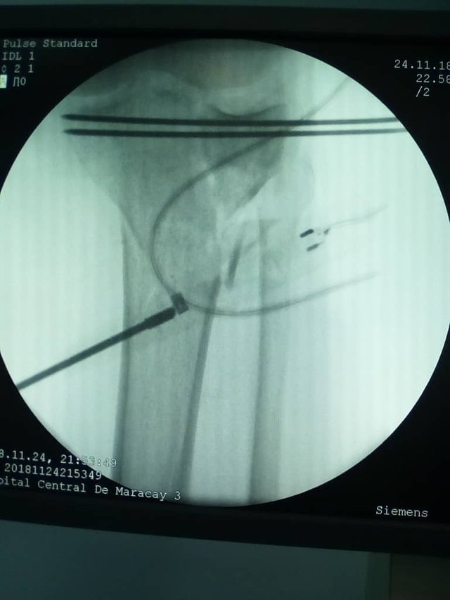
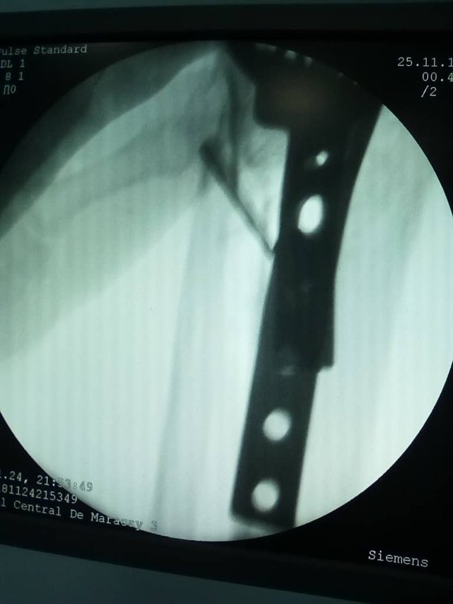
These fractures are accompanied by a high incidence of meniscus injury on the same side, especially in displaced fractures. Due to this high incidence of meniscal injury, these patients should be studied before and during the surgical procedure by means of Nuclear Magnetic Resonance and/or Arthroscopy, since if the meniscus is interposed in the focus of the fracture, an open reduction of the fracture is indicated. On the other hand, if the meniscus is intact and is not interposed in the fracture focus, the reduction is indicated closed by percutaneous fixation with cannulated or non-cannulated screws assisting the reduction by means of arthroscopy or C-arm.
Type 2- Displaced and Depressed Fractures.
In this case an adequate pre-operative evaluation is also necessary to determine the degree and location of the sinking, which may be anterior or central.
The open or closed reduction of the articular surface also depends on the state of the meniscus. The meniscus must be saved at all costs for various reasons, it helps to distribute and transmit the joint loads, it helps as a roof of the articular surface and prevents secondary displacement of the articular surface.
A lateral approach is made by which the articular surface is elevated with an awakener or impactor, then bone graft is placed to maintain the reduction, this procedure can be facilitated by the use of distractors.
When the lateral condyle is intact or with slight comminution sponge screws can be used with or without washers. On the other hand, if the comminution is very marked and the bone is osteoporotic then the use of AO plates is indicated.
Type 3- Depressed Fractures of the Articular Surface.
We must remember that this fracture generally occurs in elderly patients with osteoporotic bone after a mechanism of axial compression and valgus. If the area of the comminution is small and the joint remains stable, treatment is conservative. On the other hand if this fracture occurs in a patient with an active lifestyle then surgical treatment is indicated. The surgical treatment consists of lifting the articular surface, placing bone graft and perform crude reduction and osteosynthesis, assisted by arthroscopy and C-arm.
Type IV. Fractures of the medial condyle.
Because they are fractures caused by high energy trauma, they are generally associated with other injuries such as dislocation of the knee and neurovascular damage. These fractures usually include the tibial eminence or spine.
Conservative treatment in this type of fracture is only indicated in non-displaced fractures, as even those with minimal displacement viciously consolidate into varus.
Due to the large biomechanical forces transmitted through the medial plate, screw fixation alone is not sufficient and the use of plates is even more necessary if the lower portion of the metaphysis presents with comminution or loss of bone substance. In case of avulsion of the tibial eminence, it must be fixed with screws. In case of posterior displacement, a second incision is necessary to facilitate the reduction.
Types V and VI.
These fractures have several aspects in common, the first is that they affect the two tibial saucers and are also accompanied by a large number of associated injuries both local and general as the presence of open fractures and participation in the fracture of the tibial eminence. Type V fractures are described by Schatzker as inverted Y fractures, the fracture begins in the intercondylar area and leads to the proximal metaphysis of the tibia, separating the medial from the lateral condyle. The configuration usually consists of a displaced fracture of the medial condyle associated with a depressed fracture of the articular surface or displaced of the lateral tibial plate.
Type VI fractures differ from the previous one by their extension towards the diaphysis causing the dissociation characteristic of metaphysis - diaphysis.
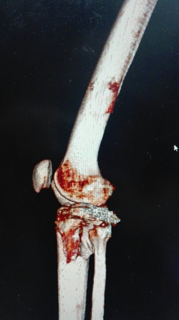
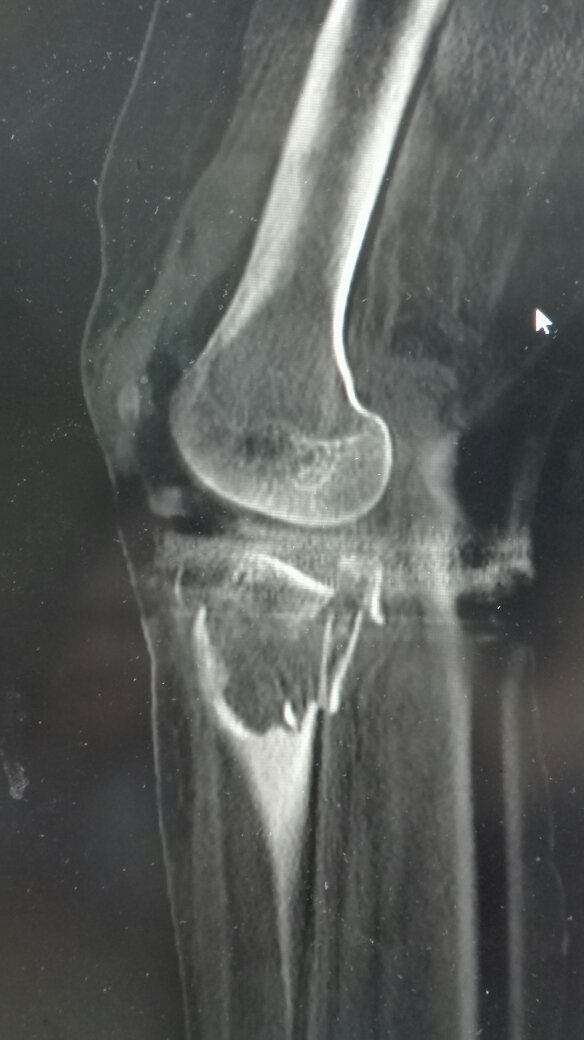
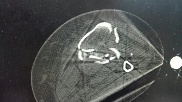
Due to the fact that they are extremely conminutive fractures, their true extension can only be determined by traction radiographs, Computerized Axial Tomography or Nuclear Magnetic Resonance.
Although the ideal treatment for these fractures is surgical, it is not free of complications. In ancient times, two AO plates were placed one on each side, which required a great dissection of the soft tissues, especially on the medial side. This brings with it a high incidence of soft tissue lesions, among which the most common are infection and wound dehydration.
In all patients the medial tibial plate fixation is of extreme importance since the most common residual deformity found in these patients is varus.
Due to the high incidence of soft-tissue complications, today a large number of authors prefer the use of external fixation with minimal approaches to the fractured area. This method allows the stabilization, reduction and a quick incorporation of the patient to the rehabilitation.
If you need recommendations or help in orthopedic surgery and traumatology do not hesitate to contact me.
Dr. Leopoldo Maizo - Orthopedic Surgeon
If you want to read more I invite you to visit my page:
Firma diseñada por @themonkeyzuelans, contáctalos vía Discord "themonkeyzuelans#9087"
Great projects from the Steemit community:

- My Fundition campaign: https://fundition.io/#!/@drmaizo/6f88ggj8h


.png)
Congratulations! Your post has been selected as a daily Steemit truffle! It is listed on rank 15 of all contributions awarded today. You can find the TOP DAILY TRUFFLE PICKS HERE.
I upvoted your contribution because to my mind your post is at least 9 SBD worth and should receive 103 votes. It's now up to the lovely Steemit community to make this come true.
I am
TrufflePig, an Artificial Intelligence Bot that helps minnows and content curators using Machine Learning. If you are curious how I select content, you can find an explanation here!Have a nice day and sincerely yours,

TrufflePigThank you very Much Friends :D