Distal radio fractures, their explanation from anatomy to treatment
Greetings friends Steemians,
Today is a pleasure to present a topic that brings a lot of discussion in the medical journals of traumatology, not only because of the diversity of information that can be found through this subject so studied, but it is our daily bread in the room emergency of a hospital in the area of traumatology.
It had always been of great curiosity to know how the fractures of children were cured, it was always something so fast, and in the end they ended up happy with a plaster. Wanting to be a traumatologist was not an idea that came to me overnight, since I stepped into the hospital during medical school I was looking to enter the traumatology area. Until I was too lucky to see when a broken wrist came along.
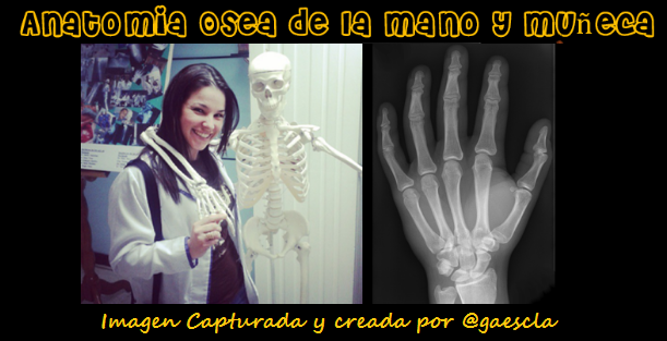
NOTIONS OF ANATOMY OF THE DOLL.
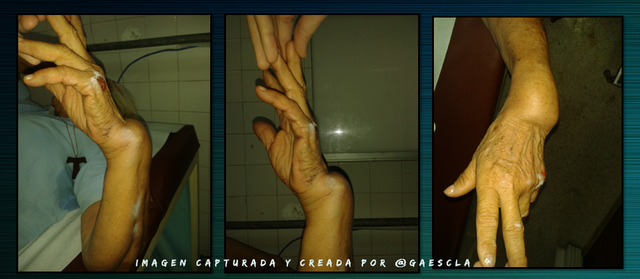
The wrist is the part of the upper limb that is going to join the hand with the forearm, and this will be joined by bones and tendons; It is important to know the bones that the wrist brings and how it articulates with the bones of the forearm.
If we see the bony part of the hand is much more illustrative for all of us, we have the fingers of the hand that are made up of 3 phalanges with the exception of the thumb that only has 2 phalanges, these in turn are articulated with the bones that are find in the palm of the hand: called metatarsals, and if we go more proximally we reach the wrist that is made up of 8 bones called carpus, these are arranged in a cross section in 2 rows with 4 bones: each of them have names : we must always name them in a coordinated order, for example from lateral to medial, the most proximal row that articulates with the bones of the forearm are: scaphoid, semilular, pyramidal, pisiform. The second row more distal and articulates with the metatasianos are: Trapeze, trapezoid, big and hamate bone.
The forearm is well known for having 2 bones inside it: the Radio and the cube (also called ulna). The radius is the one that is going to articulate with the first row of the bones of the wrist.
Here I present to my friend Felito he has always been happy and willing to teach us everything about him:
BROKEN WRIST
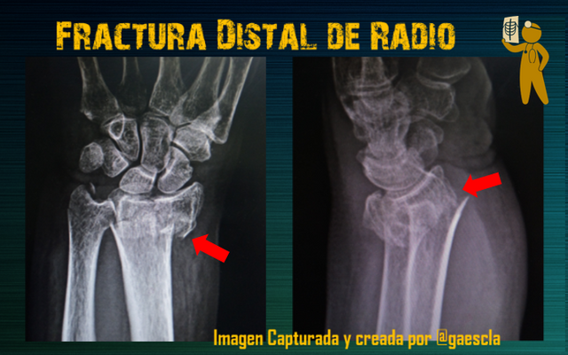
When we refer to fractures of the wrist, the most frequent are those that occur at the distal end of the radius. For this I want to present a real life clinical case.
In theory and in practice it is important to have extensive knowledge of anatomy to be able to classify a fracture in order to define its prognosis and treatment.
Arriving at the Emergency
Arriving at the emergency is always a chaotic moment, but as we are taught in medical school and in any rescue course the main thing is to keep calm, although we doctors always start all our cases with what we call current illness, really this we did it at the end of having all the data and we sat down calmly to write it.
Current disease: a 72-year-old female patient who according to the current onset of illness on 02/07/18 after falling of her own feet with upper right limb in extension and wrist in dorsiflexion, presented pain, edema, bone crackle deformity in the right wrist with limitation for its function, it goes to a medical assistance center where the clinical signs and images are evaluated, presenting a 1/3 distal fracture of the right radius.
Evaluating the RX
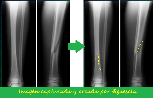
It is important when requesting an image study as x-rays should always be evaluated 2 projections, since this will give us an image in 2 different planes, allowing the doctor to know how our fracture behaves. Also in fractures of the wrist you should always see all the bones of the hand and as much as possible see more than 2/3 of the forearm, in case of a simple fracture.
Here we can see a clear example of what to see 2 projections is very important, where in the anteroposterior view no fracture is seen but in the lateral view yes. (X-rays of the left leg). Law of life we must always be very observant, but not only see one side, many can be a surprise.
For any bone deformity, the RX study is the study of choice and should always be done before manipulating any fracture. Although the clinic always prevails in the case of fractures, it is always important to document. One of the Golden rules of radiology is to make projections an articulation above and an articulation below the deformity: IMPORTANT FACT.
Studying + Our Patient
For everything there is time, we must always be calm in any medical situation, and highlight our detective skills so that all the decisions we make with our patient are correct. After knowing the current illness of our patient, and having before our eyes the diagnosis, we must focus on the antecedents of our patient:
Personal history:
Denies allergy to medications and basic pathologies, refers 2 apparently healthy children, living parents apparently healthy. Occupation: teacher of mathematics. Dominant hand: right.
I consider that in traumatic situations patients lose their sense of hearing, they increase their sense of sight and their ability to speak, so explaining a procedure is like an art: you have to have a lot of patience and having x-rays can better illustrate everything to be done.
We present a stretcher, where we send them to go to bed, it is indicated that you should place your arm inside a girth, once positioned our patient, it is explained that it must be pulled or carried to the site, we give the patient the option to place anesthetic local in the focus of the fracture.
CURING THE FRACTURE
We doctors must always have everything at hand, because once the reduction is made we can not lose it, depending on a series of criteria and many parameters, we decide where and how to perform the reduction maneuver. For this we have our dear gypsum room, which will depend on the hospital where we work, here I present where we made the case in this case. It is an office that is modified every time a patient arrives to pull.
MATERIALS
✅ Fixed frame: It is a stretcher that does not have wheels, because if we are going to pull, we do not want the table to move. If in Spanish there are many ways to name things, to the stretcher when the patient is in it or when we talk about performing a procedure in the we say table.
✅Cincha: This is a padded ring that is hung from a fixed tube in the traction area, in which the affected upper limb is placed to make axial traction easier.
✅ Injector, sterile gloves, local anesthetic, alcohol.
✅6 bandages made of mineral gypsum
✅Guata
✅ Gypsum jar where the water goes in a basin
✅ Negatoscope and the RX with 2 PROJECTIONS.
✅ 2 trained assistants: they can be welfare or post-traumatology residents.
✅A traumatologist 😉👍
PROCEDURE DESCRIPTION
We continue with our special case: Mrs. A. Fractured. PLAN: Reduced incruentra of the fracture plus confection of brachiopalamar plaster.
It is always important to start our procedures with "Previous standards of asepsis and antisepsis": it is nothing more than the cleaning of the fractured area and of course absolute alcohol with a gauze from the center of puncture in a centrifugal form, local anesthetic is placed in the center of the fracture where it is extremely important to know the anatomy, proceed to perform longitudinal traction maintained, reverse play the mechanism of production of the fracture prior evaluation of radiological study, maintain axial traction, place bandages of cotton wool (padding) protecting the pressure areas , make brachiopalmar plaster in 2 beats, that is, being careful not to apply pressure, slip plaster bandage with the correct limits, remove upper limb member, hold limb with 90 ° of flexion and hand to the zenith, study radiological control, corroborate satisfactory reduction and in the second time complete brachiopalmar plaster.
MOMENT OF APPLAUSE FOR THE MEDICAL HERO
After the plaster is placed, and the patient feels that everything is fine, all the relatives treat you like a hero, it is not a lie, literally like a hero, not only because of his performance of all the years we studied, but also because of our courage in performing procedures such as these as Cruel as they call it and more if the reduction is done by a Doctor as cute as the one described in this post.
PERSONALIZED MEDICAL INDICATIONS
We as doctors know that there are dangers after plaster casting, normally it is not necessary to indicate oral analgesics after this, it is to emphasize that the treatment to cure your broken bone is the plaster, and it must remain clean and dry, In addition, keeping the arm in a declining position can cause edema that can cause a lack of circulation, called compartmental syndrome, which can lead to many severe sequelae, so after all that happened, patients feel very afraid, once having this warning, they want to stay hospitalized.
Once again our face changes and we explain the ways in which they can prevent this from happening, and for this reason they must go to the 72 hours to be evaluated again by the attending physician, All this by writing it, previously noting the data of in the emergency notebook.
Always highlighting your indications, it does not matter that you repeat it many times and pass it in writing, repeat it to the patient and all those who will help this person during their medical rest:
KEEP ARM ON HIGH, MOVE FINGERS, DO NOT PAINT NAILS, DO NOT WIPE OR SCRATCH PLASTER, GO TO 72 HOURS TO RE-VALUATE.
Conclusions:
Well, dear readers, I hope you will enjoy an afternoon evaluating a patient with me, from how my curiosity to cure the fractures arose to give you a little knowledge about the complex anatomy of the doll in a didactic way, it is a way for us to talk more and more deep how to treat this type of fractures since we have some basic notions on how to approach this type of patients.
Bibliographic reference
Terry Canale S: Campbell Orthopedic Surgery-10th edition-2004-Elsevier- Spain
Rocwood & Greenn: Fractures in the adult. 2003 Reprint 2007. Editorial Marban.
Ya te di tu primer FLag. Andas robando publicaciones haciendo ver como si son tuyas.
No es la primera vez, ni la segunda, ni tampoco la tercera. Te comentan diciendote que pares y sigues.
Flags con Aji Picante y Mortadela
De ahora en adelante todo lo que publiques te le dare Flag por hacer plagio de contenido. Ya que no haces caso, pa que aprendas a las malas.
Link del post original