Muscle: The fibre of life
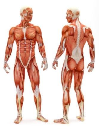
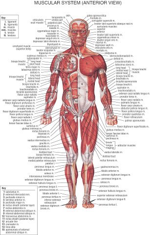
[The muscular system]
The term muscle is derived from the Latin musculus meaning "little mouse" perhaps because of the shape of certain muscles or because contracting muscles look like mice moving under the skin.
The human body consist of approximately 642 skeletal muscles and almost every muscle constitutes one part of a pair of identical bilateral muscles, found on both sides, resulting in approximately 321 pairs of muscles.
.jpg)
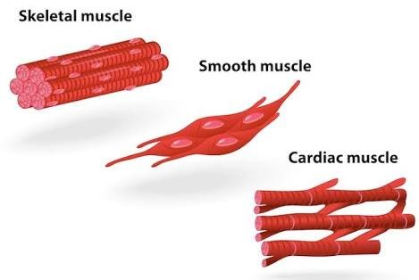
Types of muscle cells
There are 3 main types of muscle cells
- Skeletal muscle or striated
- Cardiac muscle
- Smooth muscle
Structure of the muscle
Muscle is a soft tissue found in most animals( both in humans and sub humans). Muscle cells contains a proteinous filament which is made up of Actin and Myosin that slide past one another, thus causing a contraction that changes both the length and the shape of the cell.
.jpg)
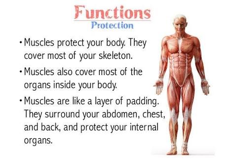
Function of muscle
The muscle helps the body perform several functions, of which are;
- They are primarily responsible for changing and maintaining posture
- They are the main catalyst of locomotion (I. E they assist in the movement of the body)
- They as well aid movement of visceral (internal) organs, such as the contraction of the heart to increase blood flow
- They aid breathing by the effective contraction of the larynx and the lungs
- They assist in peristalsis by the effective contraction of the oesophagus
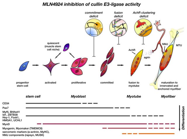
How are muscle formed
Muscle tissues are formed from the Mesodermal layer of Embryonic germ cells in a process known as Myogenesis. All muscles are derived from paraxial mesoderm. The paraxial mesoderm is divided along the embryo's length into somites, corresponding to the segmentation of the body. Each somite has 3 divisions
- Sclerotome (which forms vertebrae)
- Dermatome (which forms skin) and
- Myotome (which forms muscle).
The Myotome is divided into two sections, the;
- Epimere and
- Hypomere, which forms Epaxial and Hypaxial muscles, respectively. The only epaxial muscles in humans are the Erector spinae and small intervertebral muscles, and are innervated by the dorsal rami of the spinal nerves. All other muscles, including those of the limbs are hypaxial, and inervated by the ventral rami of the spinal nerves
During development, myoblasts (muscle progenitor cells) either remain in the somite to form muscles associated with the vertebral column or migrate out into the body to form all other muscles. Myoblast migration is preceded by the formation of connective tissue frameworks, usually formed from the somatic lateral plate mesoderm. Myoblasts follow chemical signals to the appropriate locations, where they fuse into elongate skeletal muscle cell.
Action of the muscle
Muscle action can either be classified as either Voluntary or Involuntary. Smooth and Cardiac muscle contract without conscious thought and are termed Involuntary, while the Skeletal muscles contract upon command or at will. Skeletal muscles in turn can be divided into fast and slow twitch fibers.
Muscle fuel
Muscles are predominantly powered by the oxidation of fats and carbohydrates, but anaerobic chemical reactions are also used, particularly by fast twitch fibers. These chemical reactions produce adenosine triphosphate (ATP) molecules which are used to power the movement of the myosin heads.
Anatomy of the muscle
The anatomy of muscles includes gross anatomy, which comprises all the muscles of an organism, and microanatomy, which comprises the structures of a single muscle.
Types of Tissue
The body contains three types of muscle tissue:
(a) skeletal muscle
(b) smooth muscle and
(c) cardiac muscleSkeletal muscle or "voluntary muscle" is anchored by tendons (or by aponeuroses at a few places) to bone and is used to effect skeletal movement such as locomotion and in maintaining posture. Though this postural control is generally maintained as an unconscious reflex, the muscles responsible react to conscious control like non-postural muscles. An average adult male is made up of 42% of skeletal muscle and an average adult female is made up of 36%. Skeletal (voluntary) muscle is further divided into two broad types: slow twitch and fast twitch:
Type I, slow twitch, or "red" muscle, is dense with capillaries and is rich in mitochondria and myoglobin, giving the muscle tissue its characteristic red color. It can carry more oxygen and sustain aerobic activity using fats or carbohydrates as fuel.[4] Slow twitch fibers contract for long periods of time but with little force.
Type II, fast twitch muscle, has three major subtypes (IIa, IIx, and IIb) that vary in both contractile speed and force generated. Fast twitch fibers contract quickly and powerfully but fatigue very rapidly, sustaining only short, anaerobic bursts of activity before muscle contraction becomes painful. They contribute most to muscle strength and have greater potential for increase in mass. Type IIb is anaerobic, glycolytic, "white" muscle that is least dense in mitochondria and myoglobin. In small animals (e.g., rodents) this is the major fast muscle type, explaining the pale color of their flesh.
The density of mammalian skeletal muscle tissue is about 1.06 kg/liter. This can be contrasted with the density of adipose tissue (fat), which is 0.9196 kg/liter. This makes muscle tissue approximately 15% denser than fat tissue.
Smooth muscle or "involuntary muscle" is found within the walls of organs and structures such as the esophagus, stomach, intestines, bronchi, uterus, urethra, bladder, blood vessels, and the arrector pili in the skin (in which it controls erection of body hair). Unlike skeletal muscle, smooth muscle is not under conscious control.
Cardiac muscle (myocardium), is also an "involuntary muscle" but is more akin in structure to skeletal muscle, and is found only in the heart.
Cardiac and skeletal muscles are "striated" in that they contain sarcomeres that are packed into highly regular arrangements of bundles; the myofibrils of smooth muscle cells are not arranged in sarcomeres and so are not striated. While the sarcomeres in skeletal muscles are arranged in regular, parallel bundles, cardiac muscle sarcomeres connect at branching, irregular angles (called intercalated discs). Striated muscle contracts and relaxes in short, intense bursts, whereas smooth muscle sustains longer or even near-permanent contractions.
Skeletal muscles are sheathed by a tough layer of connective tissue called the epimysium. The epimysium anchors muscle tissue to tendons at each end, where the epimysium becomes thicker and collagenous. It also protects muscles from friction against other muscles and bones. Within the epimysium are multiple bundles called fascicles, each of which contains 10 to 100 or more muscle fibers collectively sheathed by a perimysium. Besides surrounding each fascicle, the perimysium is a pathway for nerves and the flow of blood within the muscle. The threadlike muscle fibers are the individual muscle cells (myocytes), and each cell is encased within its own endomysium of collagen fibers. Thus, the overall muscle consists of fibers (cells) that are bundled into fascicles, which are themselves grouped together to form muscles. At each level of bundling, a collagenous membrane surrounds the bundle, and these membranes support muscle function both by resisting passive stretching of the tissue and by distributing forces applied to the muscle.[10] Scattered throughout the muscles are muscle spindles that provide sensory feedback information to the central nervous system. (This grouping structure is analogous to the organization of nerves which uses epineurium, perineurium, and endoneurium).
This same bundles-within-bundles structure is replicated within the muscle cells. Within the cells of the muscle are myofibrils, which themselves are bundles of protein filaments. The term "myofibril" should not be confused with "myofiber", which is a simply another name for a muscle cell.
Myofibrils are complex strands of several kinds of protein filaments organized together into repeating units called sarcomeres. The striated appearance of both skeletal and cardiac muscle results from the regular pattern of sarcomeres within their cells. Although both of these types of muscle contain sarcomeres, the fibers in cardiac muscle are typically branched to form a network. Cardiac muscle fibers are interconnected by intercalated discs, giving that tissue the appearance of a syncytium.

Ways of improving the health status of muscle cells
Physical exercise
Exercise is often recommended as a means of improving motor skills, fitness, muscle and bone strength, and joint function. Exercise has several effects upon muscles, connective tissue, bone, and the nerves that stimulate the muscles. One such effect is muscle hypertrophy, an increase in size. This is used in bodybuilding.Various exercises require a predominance of certain muscle fiber utilization over another. Aerobic exercise involves long, low levels of exertion in which the muscles are used at well below their maximal contraction strength for long periods of time (the most classic example being the marathon). Aerobic events, which rely primarily on the aerobic (with oxygen) system, use a higher percentage of Type I (or slow-twitch) muscle fibers, consume a mixture of fat, protein and carbohydrates for energy, consume large amounts of oxygen and produce little lactic acid.
Anaerobic exercise involves short bursts of higher intensity contractions at a much greater percentage of their maximum contraction strength. Examples of anaerobic exercise include sprinting and weight lifting. The anaerobic energy delivery system uses predominantly Type II or fast-twitch muscle fibers, relies mainly on ATP or glucose for fuel, consumes relatively little oxygen, protein and fat, produces large amounts of lactic acid and can not be sustained for as long a period as aerobic exercise. Many exercises are partially aerobic and partially anaerobic; for example, soccer and rock climbing involve a combination of both.The presence of lactic acid has an inhibitory effect on ATP generation within the muscle; though not producing fatigue, it can inhibit or even stop performance if the intracellular concentration becomes too high. However, long-term training causes neovascularization within the muscle, increasing the ability to move waste products out of the muscles and maintain contraction. Once moved out of muscles with high concentrations within the sarcomere, lactic acid can be used by other muscles or body tissues as a source of energy, or transported to the liver where it is converted back to pyruvate. In addition to increasing the level of lactic acid, strenuous exercise causes the loss of potassium ions in muscle and causing an increase in potassium ion concentrations close to the muscle fibres, in the interstitium. Acidification by lactic acid may allow recovery of force so that acidosis may protect against fatigue rather than being a cause of fatigue.
- Healthy diets
Eating of healthy diet or food rich in essential nutrients needed by the body tends to improve the state of well being of the muscle.
Clinical application of the muscle
- Muscle Hypertrophy
The increase in muscle size due to an increase in individual muscle cell. Independent of strength and performance measures, muscles can be induced to grow larger by a number of factors, including hormone signaling, developmental factors, strength training, and disease. Contrary to popular belief, the number of muscle fibres cannot be increased through exercise, instead muscles grow larger through a combination of muscle cell growth as new protein filaments are added along with additional mass provided by undifferentiated satellite cells alongside the existing muscle cells.
Biological factors such as age and hormone levels can affect muscle hypertrophy. During puberty in males, hypertrophy occurs at an accelerated rate as the levels of growth-stimulating hormones produced by the body increase. Natural hypertrophy normally stops at full growth in the late teens. As testosterone is one of the body's major growth hormones, on average, men find hypertrophy much easier to achieve than women. Taking additional testosterone or other anabolic steroids will increase muscular hypertrophy
- Muscle Atrophy
Inactivity and starvation in mammals lead to atrophy of skeletal muscle, a decrease in muscle mass that may be accompanied by a smaller number and size of the muscle cells as well as lower protein content. Muscle atrophy may also result from the natural aging process or from disease.
In humans, prolonged periods of immobilization, as in the cases of bed rest or astronauts flying in space, are known to result in muscle weakening and atrophy. Atrophy is of particular interest to the manned spaceflight community, because the weightlessness experienced in spaceflight results is a loss of as much as 30% of mass in some muscles.
During aging, there is a gradual decrease in the ability to maintain skeletal muscle function and mass, known as sarcopenia. The exact cause of sarcopenia is unknown, but it may be due to a combination of the gradual failure in the "satellite cells" that help to regenerate skeletal muscle fibers, and a decrease in sensitivity to or the availability of critical secreted growth factors that are necessary to maintain muscle mass and satellite cell survival. Sarcopenia is a normal aspect of aging, and is not actually a disease state yet can be linked to many injuries in the elderly population as well as decreasing quality of life.
There are also many diseases and conditions that cause muscle atrophy. Examples include Cancer and AIDS, which induce a body wasting syndrome called Cachexia. Other syndromes or conditions that can induce skeletal muscle atrophy are congestive heart disease and some diseases of the liver.
- Muscle Dystrophy
In muscular dystrophy, the affected tissues become disorganized and the concentration of dystrophin (green) is greatly reduced. - Neuromuscular Disease
Neuromuscular diseases are those that affect the muscles and/or their nervous control. In general, problems with nervous control can cause spasticity or paralysis, depending on the location and nature of the problem. A large proportion of neurological disorders, ranging from cerebrovascular accident (stroke) and Parkinson's disease to Creutzfeldt–Jakob disease, can lead to problems with movement or motor coordination.
In conclusion muscle is known to be invaluable throughout human life, as it plays several cogent roles in body compactness, so it is safe to say without the muscles, the body cannot survive.