Neurotransmission and Neurotoxins #1
With this article I provide a general insight into the function of the biochemical communication method, only animals are capable of: The Neurotransmission.
After the necessary background information is presented, selected Neurotoxins and their target points will be discussed.
Neurotransmission and Neurotoxins I

Basically, there are two possibilities in biochemical communication, namely the direct intercellular signal transmission via chemical messengers - called hormones - or a communication by electrochemical impulses. The latter is called neurotransmission, and in contrast to the former option, only higher animals are capable of doing so.
Basics on the electrochemical Signal Transmission
Although all cells have a membrane potential, only nerve and muscle cells are able to generate large changes in their membrane potential. Therefore they are collectively called excitable cells. Under resting conditions the unexcited cells have a resting potential of about - 70 mV. In response to an electrical stimulus voltage-gated channels (more on them later) are opened whereupon the membrane potential becomes more positive. This process is called depolarisation. As soon as a certain treshold is reached, a massive depolarisation is triggered. This results in an action potential, what is the actual physical nerve impulse.
For a better understanding of this, the time course of a single action potential is discussed:
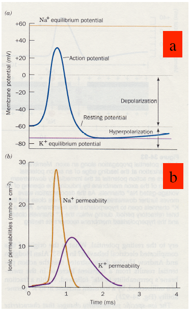
Normally the concentration of sodium is low within the cell and high in the extracellular space. The opposite is true for potassium.
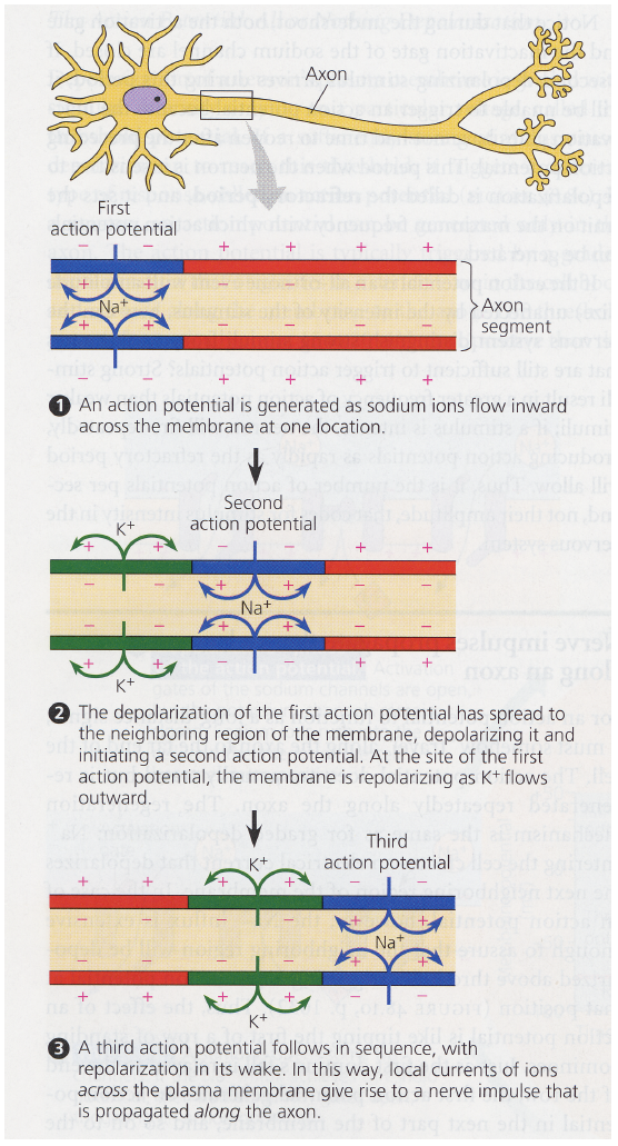
An action potential is the result of rapid permeability changes for sodium. This is shown as the orange curve in b. This rapid influx of sodium into the cell causes a depolarisation, what gives a more positive membrane potential. (Blue curve in a.) Within an array of neighboring nerve cells - e.g. an axon - the created action potential triggers a depolarisation in the neighboring membranes, i.e. the action potential propagates and the sodium channels are closing again to restore the resting potential for the initial cell.
This process is accompanied by potassium pumping in the reverse direction, hence to the outside of the cell.
As depicted in the purple curve in b.
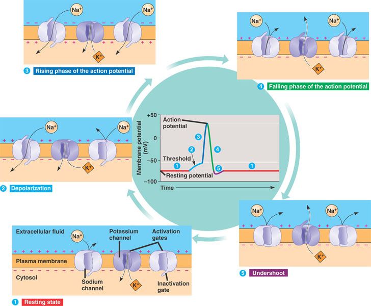
The following image illustrates the role of the voltage-gated ion channels in this process: Since the ionic radii of sodium and potassium are 0.95 Å and 1.33 Å respectively, the permeability difference between them is 100-fold! Furthermore the potassium channel needs a selectivity filter to prevent the diffusion of the smaller sodium ions through this channel.
Potassium ions are stabilized in a central cavity within the potassium channel, then they are dehydrated and tightly bound to an invariant motif (TYGYG motif) of the particular voltage-gated channel. Only ions without any hydration shell are then further allowed to cross the channel. This is how the selectivity filter works.
Propagation of the Nerve Impulse
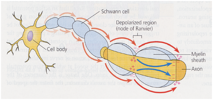
The following illustration shows how the action potential propagates along a long, slender projection of a nerve cell, called the axon.
Saltatory Conduction
Depending on the axon's diameter a nerve impulse travels at a speed of several cm/s up 100 m/s in certain invertebrates. A further increase in speed is achieved by insulating the axon with a myelin sheath deposited by Schwann cells. However, myelination is discontinuous, leaving gaps, called "Nodes of Ranvier", where the axon is in contact with the extracelullar fluid. This is shown in the next illustration. The action potential does not propagate itself in the region between these nodes, rather it "leaps" from one node to the next. This phenomenon is called Saltatory Conduction. This speeds the nerve signaling up to 150 m/s!
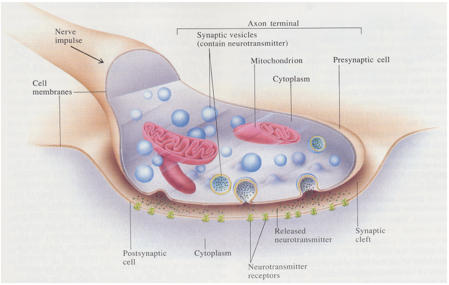
So far we discussed that a nerve cell can send an electrochemical signal down an axon, but now we need to understand how this signal is further processed as it arrives at its' destination: different nerve cells, in different locations. In principle a nerve cell sends signals down an axon, which then branches and passes the signals to the dendrites of other nerve cells at a special junction called synapses.
Astonishing additional information:
The lenght of an axon may be in the millimeter range or even go up to 1m!
The Synapse
In general there are two different types of synapses: electrical and chemical ones. Electrical synapses have a narrow synaptic cleft (20 Å) and the action potential arriving at the presynaptic membrane directly triggers a depolarisation in the postsynaptic membrane due to spatial proximity. In chemical synapses the cleft is much wider (> 200 Å) and signal transmission is coupled to the release of a special substance, called neurotransmitter, who facilitates the signal transduction via the enlarged cleft. A very typical example for such a neurotransmitter is Acetylcholin.
Function of Chemical Synapses
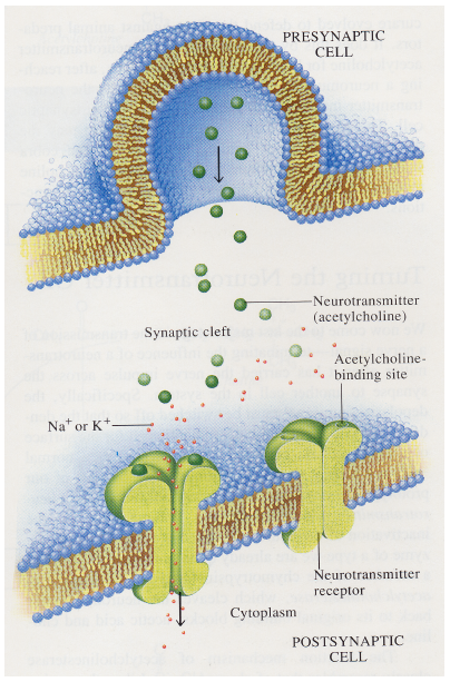
Acetylcholin is stored in presynaptic vesicles (roughly 400 Å in diameter). Each vesicle holds approximately 10.000 acetylcholin molecules. As an action potential arrives at the presynaptic membrane the opening of voltage-gated calcium channels is triggered. The resulting influx of extracellular calcium into the presynaptic terminal stimulates the exocytosis of its synaptic vesicles. This leads to a release of the neurotransmitter into the synaptic cleft. The neurotransmitter docks to the corresponding receptors of the postsynaptic membrane and leads to a stimulation in the next cell. The neurotransmitter now has to be removed from the receptor because otherwise it would give a permanent stimulus. This can be achieved via diffusion, enzymatic degradation or reabsorption into the vesicles.
With this outline all basic informations on Neurotransmission are provided and in my next upcoming article I will present some selected Neurotoxins and where they attack the shown functions of neurotransmission.
So stay tuned and follow me to learn more about interesting scientific topics!
Best,
mountain.phil28
References:
Information and non-direct-cited images are taken from
"Molecular Physiology" lectures at the TU Graz.
The only physiology I've studied is human physiology. And, it has been a while since I did that. This post brought back some old memories and served as kind of a refresher. Looking forward to more posts like this!
Congratulations @mountain.phil28, this post is the ninth most rewarded post (based on pending payouts) in the last 12 hours written by a User account holder (accounts that hold between 0.1 and 1.0 Mega Vests). The total number of posts by User account holders during this period was 3352 and the total pending payments to posts in this category was $3691.34. To see the full list of highest paid posts across all accounts categories, click here.
If you do not wish to receive these messages in future, please reply stop to this comment.
This post has received a 0.28 % upvote from @boomerang thanks to: @mountain.phil28
@boomerang distributes 100% of the SBD and up to 80% of the Curation Rewards to STEEM POWER Delegators. If you want to bid for votes or want to delegate SP please read the @boomerang whitepaper.