ASK ME PHYSICS: How is ultrasound capable of giving us 3 dimensional images
So i was asked how we get 3 dimensional images from ultrasound yet with x rays we get 2 dimensional.
Well lets start with a brief summary of x rays, the simple truth is that x rays are a high energy form of electromagnetic waves (light). The wavelength is in the region of 1 -10 nanometers. Now when they travel through material they hardly get reflected. They only get reflected by high density materials such as bone and metals. So when going through a tissue the ratio reflected is very small for each individual tissue type. So comparing those tissues becomes very hard to create depth and a 3D image.
This is made even harder by how a x ray is captured. They are/were captured on film. It is almost impossible to create a 3d x ray image on film However now with the introduction of digital x rays, 3d x rays are becoming more used however they are much more expensive.
So lets move onto how ultrasound works. Ultra sound is generally referred to sound waves over the maximum frequency of audible sound (about 20,000Hz). Diagnostic ultrasound frequencies are of the range 1-10 million hertz. Now like most forms of waves they have different ways of interacting with materials based on the structure and distribution. We can use these different interactions to picture the materials the waves are going through.
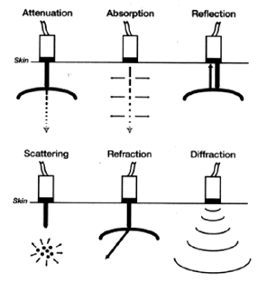
[Figure 1: Interactions of Ultrasound with tissue: Echocardiography, Bonita Anderson, Dutoit, Wiley-Blackwell]
http://www.criticalecho.com/content/tutorial-1-basic-physics-ultrasound-and-doppler-phenomenon
This picture illustrates the types of interaction these sound waves experience.
Attenuation is the general collection of the other processes. A sound wave weakens as it passes through materials as energy is lost by absorption, reflection, scattering, refraction and diffraction.
Now i can waste your time explaining what each one means, however it is not essential. The main thing we need to look at is reflection. Even though the others are important reflection is the main source of useful information we receive. A sound wave can undergo diffraction, refraction, scattering etc however if it does not reflect back to us we do not receive any information.
So a reflection (echo) occurs at the boundary between two materials. The materials have to be different enough that they have differing values of "acoustic impedance". This property is defined by the density and propagation speed of sound waves through a material. If the acoustic impedance between two materials is too similar the echo produced will be very small and the majority of the sound will continue to travel through the material. Typically a ultrasound will travel through soft tissue relatively without hindrance. We do get an echo but it is a small percentage of the overall sound pulse.
So this is the basics of why we can image in 3D. We get an echo coming back from all layers of the material. Since we do get an echo, even though it is small, from most surface interfaces we can image these interfaces. Therefore using the echo and the time for the echo to get back to us we can create a 3D image.
Here are some examples of the % of reflected pulse we get back at different interfaces
Fat to muscle = 1.08%
Soft tissue to water = 0.2%
Bone to fat = 49%
soft tissue to air = 99%
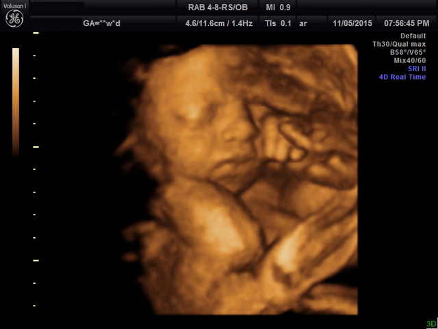
Now this should create an air of realisation. The air to soft tissue interface reflects almost all of the pulse back so how can we image anything at all? If you think to any movie you have seen of an ultrasound (eg looking at a baby in the womb) you will see that they use a gel. That is what the gel is for, it completely removes the air/tissue interface.
So there we have it.
There is an extra little bit of information on ultrasound i want to share. I found this in my research and it is rather interesting. You can image blood flow using the Doppler effect. This is where a wave hits a moving object, if the object is moving away from you then the wavelength is increased. Since the velocity of the wave propagation through the material is unchanging, then the frequency is reduced. If the object is moving towards your the wavelength is shortened and the frequency increases.
So you can measure the velocity of blood flow, which in turn means you can measure things like the size of a valve letting blood through. If the velocity of blood through a valve is very large then it follows that the pressure gradient across that valve is very large. This means that we know the valve is too small.
This is very useful as it is a safe, none invasive way of detecting potentially fatal problems in the heart and be used to identify blood clots and blockages.
Thank you for reading, any other questions please dont hesitate to ask
The Surf Graduate
I leaned something new today. Lol
Thats what im here for, any questions please dont hesitate to ask, i write posts on questions asked