GET INFORMED ABOUT YOUR KIDNEY AND ITS ANOMALIES.
Many of us including me have been hearing about kidney and its dysfunction especially, but have not known what it is all about.
So I took my time today to study and research about it and I found it interesting to share with us. we will be based solely on KIDNEY. Firstly let’s define kidney;
.jpg)
The kidneys are pair of excretory organs that are located on the posterior abdominal wall, one on each side of the vertebral column, which is behind the peritoneum. They usually excrete waste products of metabolism and excess water and salts from our body in other to maintain normal pHs.
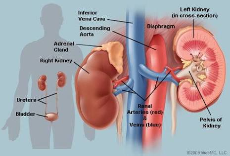
ITS LOCATION
The epigastria, hypochondriac, lumber and umbilical regions occupy the Kidney. They extend vertically from the upper border of twelfth thoracic vertebra to the center of the body of the third lumber vertebra.
Basically, the left kidney is a bit nearer to the median plane that the right, and the right one is slightly lower than the left kidney. In size, weight, shape, and orientation, each of the kidneys has about 11.5cm long, 6cm broad and 3.2cm thick.
So lets look at the blood supply. The renal arteries have blood flows in excess of close to one liter per minute. This contributes to the kidney leaving the abdominal aorta at the right angles and lie behind the pancreas and renal veins.
Concerning blood supply, each of the kidneys possesses five segments. Within the helium region, the artery typically gives rise to an anterior and also a posterior division. The anterior division creates branches that supply to the upper, apical, middle and lower segments while posterior division supplies the posterior segments.
External features of the kidney
Each kidney has a bean shape, has medial and lateral borders, upper and lower poles and also anterior and posterior surfaces.
From the anterior and posterior sides, the following structures are seen in the helium; the renal vein, renal artery and renal pelvis which expanded to the upper end of the urethra.
THE STRUCTURE
The naked eye examination of its structural coronation section shows;
An outer, reddish brown cortex
An inner pale medulla
A space In renal sinus
.jpg)
Image
The renal medulla is nearly made up of ten conical masses called the renal pyramids. The renal cortex itself is divided into two parts; the renal column which deep in between the pyramids and the cortical lobules or rather called arches which form caps over the bases of the pyramids.
The renal sinus contains the branches of the renal artery and the tributaries of the renal vein.
KIDNEY HISTOLOGY
The cortex of the kidney shows the cut section of glomeruli, many sections of proximal convoluted tubule, few collecting ducts and some section of distal convoluted tubule.
If you check through the pyramid of the medulla, it shows light staining and collecting ducts, thin and thick sections of ascending and descending limbs, capillaries and connective tissues. Wait, hope we are following...alright lets continue
Each kidney has a convex lateral margin, a concavity at the medial side that is called the helum. The helum extends into a space called renal sinus. Within the renal sinus, the pelvis divides into two or three usually called the major calyces. Each of these major calyx are subdivided into number of minor calyces that are shaped like a cup.
The kidney tissues are made up of an outer part called cortex and an inner part called medulla. This medulla consists of triangular areas of renal tissues that are called renal pyramids.
You see, these kidney that we know is deep, that’s contributes to it been expensive during any transplant due to anomaly. Let’s look at some anomalies in kidney which causes its failure.
ANOMALIES
Multisystem dysplastic kidney: it’s the one in which numerous ducts are surrounded by undifferentiated cells. Because of this, neohrons fail to develop and the uretheric buds fails to branch which causes the collective ducts not to form.
Congenital polycystic kidney disease: this kind of disease may be inherited as an autosomal dominant or recessive disorder and maybe caused by other factors.
.jpg)
Image
Autosomal recessive polycystic kidney disease (AKPKD) usually occurs within 1/5000 births, it is a progressive disorder in which cysts forms from collecting ducts. This causes the kidney to become very large and results to renal failure usually in infancy or childhood.
Renal dysplasia and agenesis: this represents primary diseases requiring dialysis and transplantation in the first years of life.
Wilmtumor: this is the cancer of the kidney that usually affects children at the age of five but may also occur in the fetus.
I believe we have learnt a lot on this wonderful and educating study, I appreciate your time. I still remain your humble friend, @angelfidel