📢 What Is Microtia? | [A Small Ear] ✔
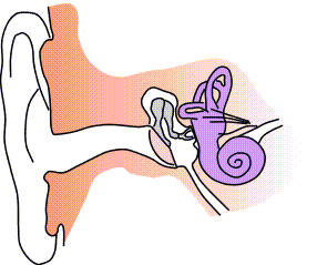
Ear Structure.Wikimedia Commons
_2.jpg)
What is Microtia?
The ear begins its development between the fifth and sixth weeks of gestation and ends at 12 weeks; therefore, the alteration occurs in this stage. Its etiology is multifactorial, including genetic factors, teratogenic, vascular anomalies and the first and second branchial arch. Likewise, it is more frequent unilaterally (70-90%), on the right side (60%) and in the male sex.
This malformation can be accompanied by other congenital defects such as: cardiac and renal, cleft palate, microphthalmia, vertebral alterations, among others. In the same way it can be part of a genetic syndrome, for example the Treacher-Collins and the velocardiofacial syndrome.
Microtia has been divided into two descriptive categories: the lobular, which is a soft mass of soft tissue, without any formation of shell or remnant of cartilage and the conchal, where there are cartilaginous portions of shell, tragus and external auditory canal.
These patients usually present with hearing loss, conductive hearing loss by 80% and therefore require studies of hearing screening, so that the child receives early care and a multidisciplinary management, which allows their insertion into school life and social in a normal way.
The reconstructive surgery of microtia is an arduous task, because it is a three-dimensional structure, with multiple anatomical repairs. This procedure is usually performed in two or four surgical times (depending on the technique and the material used). For this, the patient should be expected to be at least 7 years of age, since at this age the ear reaches adult size; although it is true that there are discrete changes in its dimensions of height and width, until reaching adulthood.

What are the different types of Microtia?
Microtia occurs in many different variations, ranging from just a small ear to complete absence of the ear, called anotia meaning "no ear." In some cases, the ear canal is very small (phonetic stenosis) or absent (aural atresia).
Several systems have been developed to describe the different types of microtia. One of the most used systems to categorize the different "grades" is illustrated below:
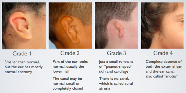
Microtia.Wikimedia Commons
What causes microtia?
The cause of microtia is not well understood, particularly on the role of environmental and genetic factors. Genetics are believed to be a cause in only 5% of all patients. Several theories have been proposed to explain the cause of Microtia during fetal development, such as cells of the neural crest disturbances, vascular disruption, and altitude, but these have not been demonstrated.
It is important to understand that nothing a mother does during pregnancy, such as coffee drinking, alcohol consumption, or even drug abuse, has been shown to cause Microtia. However, when taken in the first trimester of pregnancy, some medications have been linked to microtia, including thalidomide and Accutane.
Can Microtia involve more than one ear?
Microtia usually occurs on only one side (more frequently on the right), but 10% of patients have microtia on both sides (bilateral microtia).
Microtia is often seen as an isolated entity, but it can also occur with other syndromes such as hemifacial microsomia, Goldenhar syndrome, or Treacher Collins syndrome. Other syndromes with microtia can also affect the kidneys, heart, eyes, craniofacial bones, and the skeletal system. These children are usually looked after by a Craniofacial Team.
Auditory test and Image Test
Auditory and imaging studies, mainly tomography, are among the studies that the patient requires. The auditory exams include: otoacoustic emissions, brainstem evoked potentials, free field audiometry, tonal and vocal audiometry and impedance or tympanometry, among others. The choice of evaluations will depend on the age of the patient and the clinical findings.
Discussion
Microtia is a malformation of the auricular pavilion, which may or may not be accompanied by any other syndromic manifestation. The most common is that it is unilateral and right, as it happens in the majority of patients.
It is important to rule out in these patients alterations in other organs and systems, initially renal, due to the relationship between malformations of the auricular and renal pavilions. On the other hand, screening for hearing loss should be done, not only to reveal anomalies in the otic structures, but also to initiate early stimulation early.
It is also important to point out that the psychological support of the parents, of the family and school environment is vital, in order to avoid the child being mocked and not underestimating the impact on any human being, the fact of looking different.

References
- http://microtiaearsurgery.com/
- Aguinagua-Ríos, M. (2014). Microtia-atresia: clinical, genetic and genomic aspects. Medical Bulletin of the Children's Hospital of Mexico. Vol 7. Nº6. P.p. 387-395.
- Sorolla, Juan y col. (2012). Current management of microtia: Anatomo-surgical redefinition. Rev Chil Cir vol 64. Nº 6. P.p. 528-534.
Imagen Sources: All images meet the requirements to be published.
|| @utopian-io || @anomadsoul || @acidyo || @ocd-resteem || @steemstem || @vortac || @cervantes || @hendrikdegrote || ¡Vote for witness! Here: https://steemit.com/~witnesses


That paragraph is duplicated in the post.
Done