Viral Hemorrhagic Fevers: Killer Diseases that turn humans into Biological Weapons
Introduction
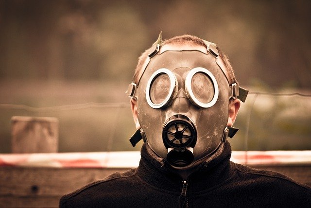 The Ebola Outbreak was a nightmare to mankind (License: Public Domain]:
Pixabay
The Ebola Outbreak was a nightmare to mankind (License: Public Domain]:
Pixabay Between the years 2013 and 2015, Africa suffered the biggest outbreak of Ebola virus disease ever recorded in history. The disease spread through West Africa, affecting countries like Guinea, Sierra Leone, Liberia, Senegal, Mali and Nigeria, infecting more than 28,000 people, and leaving at least 11,000 dead.
Ebola virus, just like Zika virus, Lassa Fever, etc, is one of the group of diseases called Hemorrhagic Fevers, which are very contagious and can spread like wildfire in any geographical area.
Viral Hemorrhagic Fevers are different groups of animal and human diseases caused by four families of RNA viruses namely Filoviridae, Flaviviridae, Bunyaviridae and Arenaviridae. All hemorrhagic fevers manifest with symptoms such as fever and bleeding disorders, but the most severe ones like Ebola, Lassa fever and Marburg virus cause lethal bleeding, loss of organ coordination and death.
To better understand the different kinds of Hemorrhagic Fevers, we'll discuss them according to their causative organisms.
Causes of Hemorrhagic Fevers
Hemorrhagic Fevers are caused by four groups of zoonotic RNA viruses. (Zoonotic viruses are viruses which naturally live in animal hosts). They include
- Arenaviridae (Arena viruses)
This family of viruses live mainly in different species of rodents. The viral particles are spherical in shape with a very tiny diameter of 110 to 130 nanometers enclosed in a lipid membrane.
The viral particle is made completely of only RNA, and although their replication system is yet to be totally understood, it has been established that their new virion particles are formed by budding through the surface of their host cells. Examples of arenaviruses are Junin virus, Machupo virus Lassa virus which causes Lassa fever.
- Filoviridae (Filoviruses)
Filoviruses are pleomorphic in nature (They can exist in different shapes). Some have long (up to 14,000 nanometers) branched filaments of circular, “U” or “6” shape enclosed in a lipid membrane. The virion particles contain a single molecule of single-stranded negative-sense RNA. Just like arenaviruses, their replication system is not fully understood yet, but they replicate by budding out of the surface of the host cells.
Only two Filoviruses have been identified so far: Ebola virus and Marburg virus.
- Bunyaviridae (Bunyaviruses)
They're also single-stranded negative sense RNA viruses enveloped in a lipid membrane. They're found in arthropods (ticks, mosquitoes and sandfly) and rodents. Examples include Hantavirus and Crimea-Congo hemorrhagic fevers virus.
- Flaviviridae (Flaviviruses)
The yellow fever virus belongs to this family. Flaviviruses have diameters of about 40 to 60 nm. They're positive-sense single stranded RNA viruses of spherical and icosahedral geometry, enveloped in a lipid membrane. They cause diseases such as hepatitis encephalitis and a birth defect called microcephaly.
Having established a background, now let's talk about two of the most endemic Hemorrhagic Fevers that has affected Africa in recent times: Ebola virus fever and Lassa virus fever.
Ebola Virus Disease
Ebola fever is a viral haemorrhagic disease which affects humans and other primates. It is caused by the Ebola virus, and its symptoms include high grade fever, muscular pain, severe bleeding, sore throat, headache and diarrhea, most of which begin to manifest from 2 days to 3 weeks of contracting the disease.
Other complications include low blood pressure due to excessive loss of fluids, decrease in kidney and liver function and life threatening internal and external bleeding. Ebola fever has a high mortality rate, Ebola virus is spread by direct contact with the body fluids of people who already have the disease, or by touching or eating infected animals like bats and monkeys.
Structure of Ebola Virus
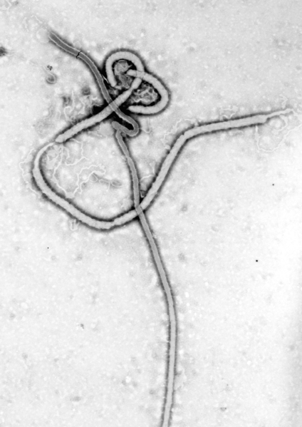 The Ebola Virus (License: Public Domain]:
Wikicommons
The Ebola Virus (License: Public Domain]:
Wikicommons 
The Ebola virus belongs to the filoviridae family. There are five strains of Ebola virus (Zaire, Bundibugyo, Sudan, Reston and Tai forest), but amongst them all, Ebola Zaire strain is most virulent.
Structurally, the Ebola virus looks like a thread of approximately 970 nm length and 80 nm diameter. It mostly exists in long filamentous and cylindrical body, and can sometimes come in a “U” shape, circular shape or “6” shape. Ebola virus has viral glycoprotein spikes of 7-10 nm projecting from its outer viral envelope (which is derived from the host cell membrane).
The viral envelope contains the nucleocapsid which consists of a single stranded RNA genome wound around the nucleoprotein (np), polymerase complex protein (VP30), minor nucleoprotein (VP30) and L protein.
Mechanism of infection of Ebola Virus
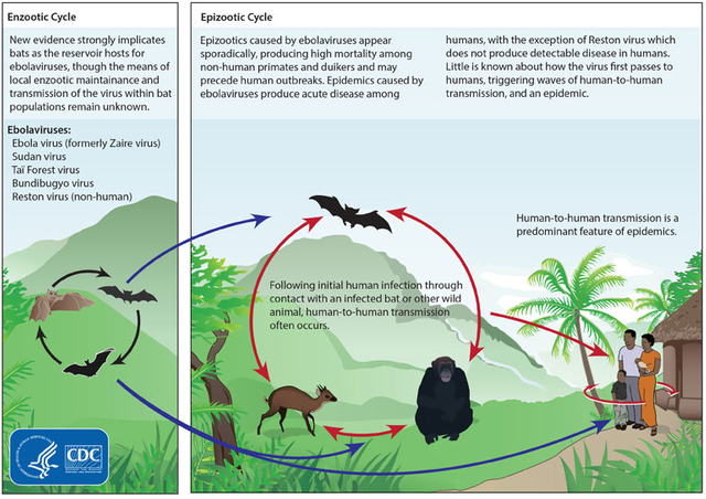 Mode of Transmission of The Ebola Virus (License: Public Domain]:
Wikicommons
Mode of Transmission of The Ebola Virus (License: Public Domain]:
Wikicommons 
The mechanism of infection of Ebola virus is poorly understood, but it is well known that the virus penetrates the human body through abrasions, mucosal surface, injuries on the skin or directly through the skin surface when an uninfected person comes in contact with the body fluids of an infected human or animal.
Ebola virus thus invade the cells using mechanisms such as macropinocytosis and receptor mediated endocytosis mediated by through the glycoprotein. The virus is delivered into the acidic endosome of the cell, where the viral envelope is uncoated to release the genomic RNA into the cytoplasm.
The most interesting thing about the Ebola virus is that it has a way of totally evading the immune system, such that it can go ahead and wreck havoc while the body immune system would still be busy trying to find out what the problem is.
As soon as the Ebola virus makes its way into the body, it targets the cells of the immune system. It's first targets are the dendritic cells. The dendritic cells can be described as the signaling cells of the immune system. They normally show signals of an invading infection and cause the activation of T-lymphocytes which destroy infected cell before the pathogen can replicate further.
Once the Ebola virus successfully destroys the dendritic cells, they would no longer be able to present signals for the activation of T-cells, and antibodies will not be formed to combat the infection. The virus therefore takes advantage of the defective immune system to replicate rapidly using the host's cells.
Once a person is infected by this disease, he/she becomes a walking biological weapon because it takes only a single contact with a healthy person to place that person on the same “death row”.
Symptoms of Ebola Virus
Bleeding from the nose, gastrointestinal tract, guns and other internal and external surfaces.
Anuria (Inability to form urine)
Impaired blood clotting
Bloody vomit, reddening eyes
Multiple organ dysfunction which results in death.
Treatment and Prevention
Till date, there has been no established cure for Ebola fever. It has over 90% mortality rate, and the only way to prevent it is by avoiding contact with body fluids and corpse of people or animals infected with the disease.
Lassa Fever
Lassa fever is a fatal haemorrhagic fever caused by the Lassa virus. The Lassa virus belongs to the Arenaviridae family of viruses, and its main host is the rodent Mastomys natalensis (aka multimammate mouse). It was first reported in Nigeria,West Africa in 1969.
Lassa fever is not as contagious as Ebola virus fever as it is not normally passed from person to person, instead, Humans typically get infected by the disease through the urine or feces of mastomys rats. In rare cases, health workers might get infected through direct contact with the blood and body fluids of infected patients.
Structure of Lassa Virus
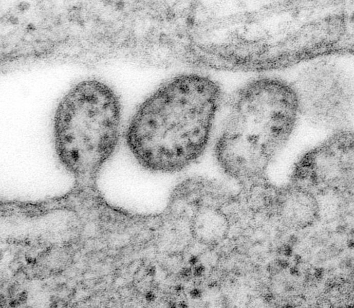 The Lassa Virus Structure (License: Public Domain]:
Wikicommons
The Lassa Virus Structure (License: Public Domain]:
Wikicommons 
The Lassa virus is pleomorphic in nature. This entails that It can exist in round, oval or any other shape. It has a diameter of 110 to 131 nm and it has an outer envelope which contains two single stranded RNA segments, the small S-segment and the large L-segment.
The L-segment encodes the viral RNA polymerase and zinc binding protein, while the S-segment encodes the precursor of the surface glycoprotein and structural protein-nucleoprotein. The surface glycoprotein precursor is then glycolytically cleaved to form the envelope glycoproteins GP1 and GP2, which facilitate the entry of the virus by binding to alpha dystroglycan receptors on the host cells.
Mechanism of Infection of Lassa Fever
Lassa virus enters the host cells through cell surface receptors, alpha dystroglycan. Some amino acids present on the GP1 protein on the surface of the virus aide high affinity binding of the virus to alpha dystroglycan. Lassa virus suppresses the immune system and replicates very quickly in the body.
Symptoms of Lassa Fever
Some symptoms of Lassa Fever include;
- High grade fever
- Depression
- Mucosal bleeding
- Facial edema
- Internal bleeding
- Weakness
- Convulsion
Treatment of Lassa fever
Currently, there has been no confirmed vaccine used for treating Lassa fever, but a drug called Ribavirin has been proven effective when administered within six days of exposure to the virus.
Conclusion
Amongst other hemorrhagic fevers, Ebola fever and Lassa fever have caused major concerns in sub Saharan Africa. Because these diseases do not have any sure cure, it is better to prevent exposure and spread of the disease. It is recommended that we report all cases of severe fever to health-care providers and avoid contact with body fluids of infected persons.
People mostly at the risk of infection are those living in rural areas where one can easily get in contact with wild animals which may be vectors of the disease, so we are strongly advised against eating Bush meat especially those sold by the roadside, and to cook all meat properly before eating. Also, maintaining a clean and healthy environment can reduce the risk of invasion of rats which may spread Lassa virus.
References
Arenaviridae. Retrieved on May 7th, 2018.
Filoviridae. Retrieved on May 7th, 2018.
MOI of the Ebola virus. Retrieved on May 8th, 2018.
Lassa Fever. Retrieved on May 9th, 2018.
Great job sir.... You did well enlightening us about this virus.
We are still grateful it didn't kill us all in Nigeria.
Hehe.... Lol. No be lie. I was so scared that I had to learn how to produce hand sanitizers. I heard it's back in some African countries now. I hope it doesn't cross our borders again.
Hehehe.. That time was horrible.
I hope it doesn't get back into the country.
Sad how we can't effectively tell the viruses to stop multiplying or to inhibit their receptors or our cells receptors like the other antivirals do.
We still have a lot of research to do in order to say that we have a cure for the main viruses.
Exactly. We have a lot of research to do, and sadly, the most affected part, Africa, has limited resources to make those research happen.