MEDICAL DIAGNOSTIC IMAGING; A LIFE SAVER!
Since it is virtually impossible to look at a sick person and figure out what is wrong, so a need for method of knowing the problem with a sick person arises. Need for medical diagnostic imaging arise. Medical imaging in a simplest term means method use in producing image of internal organ without necessarily opening up the body.
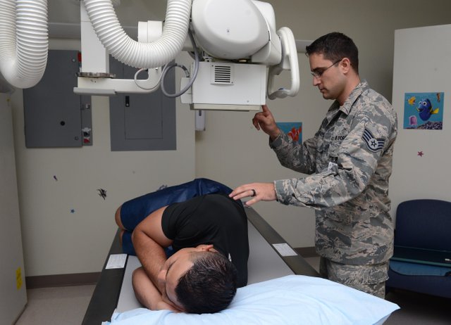
Public domain: U.S Airforce photo by Airman 1st class Arielle released
There are various forms of diagnostic imaging. X-ray, Magnetic Resonance Imaging(MRI), Ultrasonography will be discussed here.
X-radiation
Radiography is a science that deals with imaging technique. X-ray is the most common. Welhelm Rontgen discover a new type of radiation called he called X-rays. X-rays are used for making diagnosis and treatment for diseases. Diagnosis is done through viewing structure of internal organs with barest harmful effect provided all precautionary measures are taken into place. X-rays images are produced on films called radiographs. . Some organs that allows X-rays to pass through them are referred to as radiopaque example bones. Organs that do not allow X-rays to pass through them are referred to as radiolucent for example stomach. Radiopaque is also called radiodense and they are white on radiograph while radioluscent organs are black. It enables diagnosis of fracture and clinical disease like tuberculosis. X-rays can be used in specialized form of imaging like computed tomography and fluoroscopy.

How does x- ray work?
The beam travels through the air from an x-ray machine and comes in contact with body tissue(part where x-ray is to be taken) and its environment thereby producing image of the exposed part of the body on a radiograph. The film is usually placed below the exposed part of the body.
The x-ray machine produces the x-ray. The faster the electron from the source are moving the easier they penetrate the human body or other target. In an x-ray tube some sort of metals are used. X-ray continues moving in the targetted area unless they are absorbed by lead.
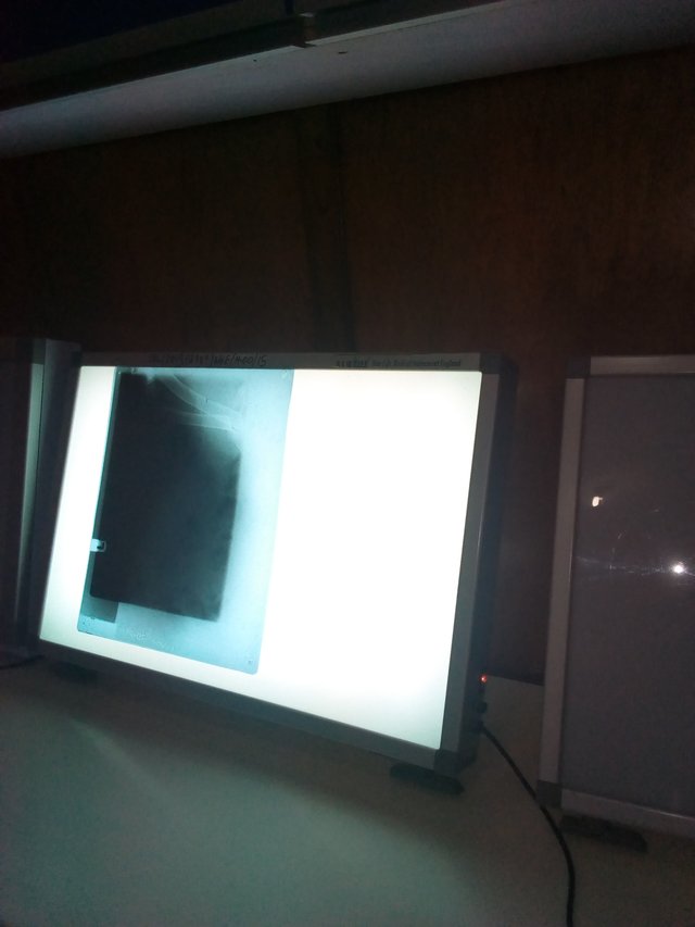
X-ray are usually directed towards the body part been examined. The rays penetrate through moving easily across different tissue of the body. Only radiodense body part like bone and teeth absorbs enough rays to be reflect on the x-ray film. The x-ray film is processed just same way photographic films are developed. The radiologist then reads the radiograph(the x-ray film) produced on the film in a light source.
It is more like negative of photographs because light and dark are formed on it. Soft tissue in the body are usually darker because they are radiolucent while bones are lighter because they absorb the x-ray.
If radiolucent areas like stomach and other soft tissue radiographs are about to be taken the doctor gives the patient a radiopaque liquid to drink and also inflate the organ well to ensure proper viewing of the soft tissue. A soft tissue filled with barium will show clearly because the barium will absorb the radiation and also the gas using to inflate the stomach will help in proper viewing of it. This barium compound is called contrast media.
Contrast media is most time use in conjunction with a fluoroscope when radiograph of radiolucent area is to be taken. In fluoroscopy x-ray passes into the body onto a fluorescent screen. This then produces a moving image. Fluoroscopy can be used to study the movement of the contrast media in the body.
As we know that technology is moving forward, it paramount to emphasis fact the several development has been made in viewing internal organs for diagnosis. Some of this development uses x-ray while some don't. But the general term medical imaging is used in describing all the various method. Medical imaging helps to give doctors image of what is inside the body enhancing diagnosis and management.
Precaution in an x-ray unit.
It's a must proper safety observed in a radiation unit. As useful as the x-ray is, it also has negative effect. The long-term effect of radiation result in dermatitis(inflammation of the skin), cell shrinking(atrophy), sterility( inability to perform), causes hair loss and also causes cancer. X radiation mostly affect actively growing cells.
Their several ways this is can be prevented. The patient and the assistant are protected. The assistant is protected because he holds patient firm and ensure no movement. The x- ray machine must also be in good condition for safety. This leads to several precautions that must be observed in an x-ray unit.
Patient safety: Safety of the patient to take X -ray is important so to achieve this the patient must wear a lead apron, a collar mostly a thyroid collar, groin pads to protect the testes, gobble to protect the eyes and some other things. The lead apron is worn to protect the whole body from absorbing x-ray from the machine.
X-ray machine: It important as well that it is a good condition for optimum safety. Also, harmful radiation must be filtered out and x rays must be restricted to the smallest possible area so as to prevent excessive escapes of radiation.
Now onto protection of the assistant, he must wear the above and also a dosimeter which is basically used for measuring the stray radiation around. An abnormally high reading of the machine is bad and effect are detrimental. Also, concerning the environment it must be lead, no windows, caution signs must be present as well.
Magnetic resonance imaging
MRI uses magnetic field and radio waves to create images that are very diagnostic. Its gives images that are clear in outline. MRI uses large magnetic, computer and radio waves. It is a non invasive( doesn't require opening) diagnostic method. Most importantly MRI does not use ionizing radiation like CT scan and x-rays which maybe potentially harmful to patient, handlers, assistant as well as people around(when it goes into the environment). MRI gives cross sections of an organ which when combined is diagnostic.
How MRI works
There are 2 magnet which forms bulk of MRI. The body is made up of water molecule which includes hydrogen and oxygen atom. In elementary physics an atom is known to contain proton in its nucleus which same applies here. The proton in this 2 atoms is very sensitive to magnetic field. Water is present all over the body, but not uniformly arranged. Upon entering an MRI, it makes the water molecule to align in a particular direction which could be north or south. The magnetic field is turned on and off very quick. This makes the hydrogen atom orientation alter and it then quickly move back to its relaxed state. The magnetic field is created by passing electricity through coils present. This is then response for the sound produced by the MRI machine when its in use. The computer involved is then used to generate an image that can be interpreted by a doctor. The images are usually 3-D images.
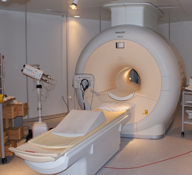
Wikimdeia creative common: creative common attribution 3.0 unported
Uses of MRI are quite enormous which I will be shedding light on. Firstly, MRI is used to study and diagnose brain and spinal cord abnormality. These are special organs involve in co-ordination of the body. It as well measure brain activity by monitoring blood flow to the brain. Secondly, help to diagnosing heart problem. There are several condition that causes severe heart problem some can be even be congenital. MRI helps in diagnosing it. Ladies who have fibroid and endometriosis are diagnosed using MRI because it as well causes severe pelvic pain in women. Not to be left out is the fact that its help in diagnosing uterine problem in women undergoing evaluation for infertility. Furthermore, tumor, cyst and other abnormalities are diagnosed too with MRI. MRI is such a useful machine in diagnosis just that it can't be afford by many hospitals in my country...lol
Precautionary measures are also important when about to take MRI. Firstly, the patient must ensure all metals substance with him are all removed. Example like jewelries, gold chains and others. Also a contrast liquid like barium is also applied here too like i made mentioned in X-rays. The barium help improves the appearance of certain body tissue. Also, patient are given headphones to reduce noise from the MRI machine which could help remove anxiety in some patient. Furthermore, all movement of the patient are restricted and staying still is important when scanning is to begin so as to avoid disruption. At any point, when the patient is uncomfortable he can assistant for help
ultrasonography
Ultrasonography is diagnostic imaging method which uses high-frequency sound waves and as well the echoes from the sound waves to create the image of the body organ or tissue. It is also called medical sonography. Just like MRI discussed above it is relatively safe and noninvasive. It doesn't use ionizing radiation like X-ray discussed earlier.
Now onto how ultrasounds work, it usually involves the use of ultrasound gel which is applied directly on the skin then a transducer(a probe) is applied on the skin surface to transmit sound waves with high frequency which is usually inaudible to the ear. The transducer gets the echoes( the sound that bounces back) and sends it to the computer. The computer uses the sound to create an image on the screen.
Importantly, ultrasound display images of the organ or tissue being examined in a flat section and thin. Also, we have 3-D ultrasound images meaning images from 3 dimensions.
Ultrasound generally consists of video screen together with the earlier mentioned part. The gel is applied to enable sound wave travel to tissue and then back. Ultrasounds are applied in several medication diagnoses which includes an examination of a baby in a pregnant woman. Also helps in diagnosing the cause of pain in the body. In addition, ultrasound helps in assessing damage when heart attack occurs. In conclusion of the uses, it also helps in diagnosing some special heart-related conditions like congestive heart failure, valvular defect. Ultrasound of the heart is especially unique and it called echocardiogram.
Lastly,
In conclusion, will like to discuss likely futures of diagnostic imaging techniques. In just an hundred years into advent of radiology as a field. It has gone so deep that it has greatly enhanced diagnosis in humans and also industrial uses. Though the advantages overweighed the disadvantages. If all this happen in just a century. What will happen in future?
Surely, progress in diagnostic imaging will continue at just a great pace. More sophisticated machine for diagnosis and treatment will be developed as well it will be safer than what we have presently.
Not to be left out, in the future there will be more linkage between computer and radiation machine thereby enhancing doctors ability to view internal organs, make diagnosis, treat and as well as study for academic purpose. Also ultrasound will become clearer and easier to interpret in the future. Just the way doctors uses stethoscope for auscultation during body examination, ultrasound will be used as such thereby making diagnosis easy.
References
- Kathy Winkler, Inventors and Invention Radiology.
- Dr Aliyu veterinary radiologist. University of Ilorin.
- Radiation safety
- How X-ray works
- What you should know about MRI scans
- General Ultrasound
- Medical Imaging Techniques: Types & Uses

If you donate 1 SBD or STEEM to @a-a-a I will resteem your last post to over 72,500 followers on my 2 accounts @a-a-a and @a-0-0
Hey @tundevet
I read this and I must tell you I am somewhat afraid now because I weren't wearing any protective cloth or whatever, when I had to be subjected to x-ray imaging following my accident. I was subjected to this imaging both in Nigeria and India without protection. Please, do I need to entertain any fear?
Regards.
@eurogee of @euronation and @steemstem communities
Same here when I had x-ray there no form of protection.
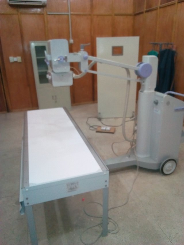
There are different type of x-ray machine some very large ones require you to put on a protectant. While some don't require especially small x-ray machine. There are some x -ray so portable that it can be used on the field. Also it can be because the part about to be expose is small and then focused by expert. @eurogee
Entertain no fear. The radiologist know the reason why you don't need. @eurogee
I see. Thanks
Congratulations! This post has been upvoted from the communal account, @minnowsupport, by tundevet from the Minnow Support Project. It's a witness project run by aggroed, ausbitbank, teamsteem, theprophet0, someguy123, neoxian, followbtcnews, and netuoso. The goal is to help Steemit grow by supporting Minnows. Please find us at the Peace, Abundance, and Liberty Network (PALnet) Discord Channel. It's a completely public and open space to all members of the Steemit community who voluntarily choose to be there.
If you would like to delegate to the Minnow Support Project you can do so by clicking on the following links: 50SP, 100SP, 250SP, 500SP, 1000SP, 5000SP.
Be sure to leave at least 50SP undelegated on your account.
Hi @tundevet!
Your post was upvoted by utopian.io in cooperation with steemstem - supporting knowledge, innovation and technological advancement on the Steem Blockchain.
Contribute to Open Source with utopian.io
Learn how to contribute on our website and join the new open source economy.
Want to chat? Join the Utopian Community on Discord https://discord.gg/h52nFrV