Japanese scientists revived the dead brain of a mouse

Tissues were able to remain alive and maintain circadian activity for 25 days.
Scientists from the Riken Center for Research on the Dynamics of Biological Systems in Japan using the new technique were able to keep the dead brain tissue of the mouse alive and functioning for almost a month. The results are published in the journal Analytical Sciences.
It used to be difficult to keep a tissue - or, for example, organs intended for transplantation - alive for more than a few days: the problem is that, on the one hand, the tissues will dry quickly and die if they are not stored in a nutrient-rich moist environment, with another - excessive placement of tissue in a liquid, drowning in it, can lead to damage, disrupting gas exchange.
During the experiment, scientists used a special microfluidic device, which helped moisturize and prevent the drying of tissues explanted from the brain of the mouse, but at the same time maintain optimal hydration.
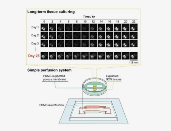
The bottom line is that the device contained a semi-permeable channel coated with an artificial membrane and walls made of polydimethylsiloxane (PDMS), a chemical often used as an antifoam agent in OTC drugs.
This means that the tissue does not need to remain constantly immersed in the liquid: it could receive nutrients from a moist environment, since it circulated in a semipermeable channel and passed through an artificial membrane without disturbing gas exchange.
“It was difficult to control the flow of the medium because the microchannel that formed between the walls of the PDMS and the porous membrane was unusual,” explained lead author Nobutoshi Ota. “Nevertheless, we succeeded after testing and modifying the porous membrane and adjusting the inlet / outlet flow rates.”
When the flow was established, the researchers tested the device on tissues from the dead mouse brain responsible for regulating the circadian rhythm - the suprachiasmatic nucleus.
The rodents used in the experiment were genetically modified so that the activity of their circadian rhythm was associated with the production of a fluorescent protein, which allowed scientists to track tissue viability through the amount of bioluminescence produced.
As the results of the study showed, tissues can remain alive for more than 25 days, maintaining circadian activity. On the 25th day, they retained approximately 97 percent of the initial activity, scientists noted. In this case, the control sample lost activity after 10 days.
“This method can be used not only for explanting animal tissues,” Ota said. “In addition, it will improve research on organogenesis.”
Now a team of scientists from Riken is focused on long-term experiments in which they are going to observe the formation of blood vessels and cell movement during the development of organoids.
What do you think about this?
Write in the comments!
Subscribe to my blog!
Vote for me!